1.2 - Muscles & Movement
The musculo-skeletal system and its role in movement
The musculo-skeletal system combines the skeleton and muscles to enable body movement. Muscles attach to bones via tendons, which are strong connective tissues that transmit force. When muscles contract, they generate tension that pulls on bones, causing movement at joints.
Key components of the musculo-skeletal system
- Skeleton - Provides a rigid framework of bones that support the body and serve as attachment points for muscles
- Muscles - Contractile tissues that produce force and movement by shortening or creating tension
- Joints - Points where bones meet, allowing controlled movement through muscle actions
- Tendons - Tough, fibrous tissues connecting muscles to bones, enabling force transfer without direct muscle-bone contact
Major muscles and their specific actions at joints
The body has several major muscles that perform specific movements at joints.
Functions and examples of major muscles
| Muscle | Location | Action and example |
|---|---|---|
| Biceps | Located in the upper arm | Causes flexion at the elbow, for example, when performing a weight curl |
| Triceps | Located in the upper arm | Causes extension at the elbow, for example, during the release phase of a basketball jump shot |
| Pectorals | Located in the chest | Cause adduction and horizontal flexion at the shoulder, for example, during a tennis forehand drive |
| Hip flexors | Located at the front of the hip | Cause flexion of the leg at the hip, for example, when lifting the knee in a sprint |
| Gluteals | Located in the buttocks | Cause extension, rotation, and abduction of the leg at the hip, for example, pushing forward during running |
| Deltoid | Located at the shoulder | Causes flexion, extension, abduction, or circumduction at the shoulder, for example, during the arm pull in front crawl swimming |
| Latissimus dorsi | Located in the back | Causes extension, adduction, or rotation at the shoulder, for example, during the pull phase of the butterfly stroke in swimming |
| Tibialis anterior | Located at the front of the lower leg | Causes dorsiflexion at the ankle, for example, during a heel-side turn in snowboarding |
| Rotator cuff | A group of muscles (including infraspinatus, teres minor, and subscapularis) around the shoulder | Causes rotation and abduction at the shoulder while stabilising the joint, for example, when lifting arms to prepare for a dive |
| Hamstrings | Located at the back of the thigh | Cause flexion at the knee, for example, drawing the foot back before kicking a football |
| Quadriceps | Located at the front of the thigh | Cause extension at the knee, for example, during a rugby drop kick |
| Gastrocnemius | Located at the back of the lower leg (calf) | Causes plantar flexion at the ankle, for example, when standing on toes in ballet |
| Abdominals | Located in the core | Cause flexion at the waist, for example, during a sit-up |
Types of muscle contractions: isometric and isotonic
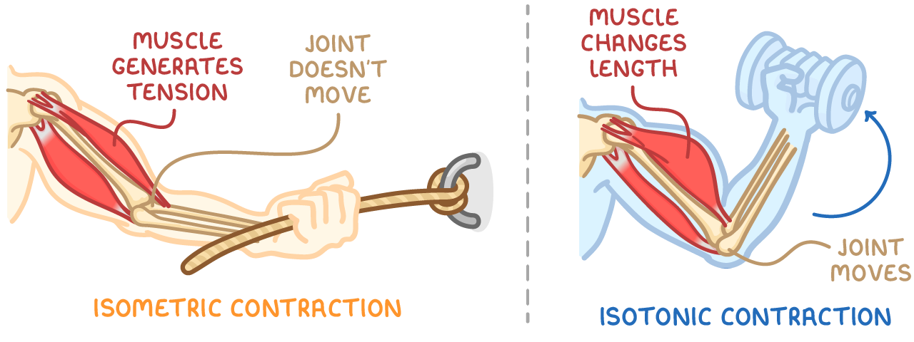
Muscle contraction occurs when muscle fibres generate tension to apply force to bones, enabling movement or stability. There are two main types: isometric and isotonic.
Isometric contraction
In an isometric contraction, the muscle generates tension but remains the same length, resulting in no movement at the joint.
For example, pulling on a fixed rope attached to a wall, where the arms and rope stay stationary despite the effort
Isotonic contraction
In an isotonic contraction, the muscle changes length while generating tension, causing movement at the joint.
For example, lifting a dumbbell, where the arm bends and the weight moves upwards
Concentric and eccentric isotonic contractions
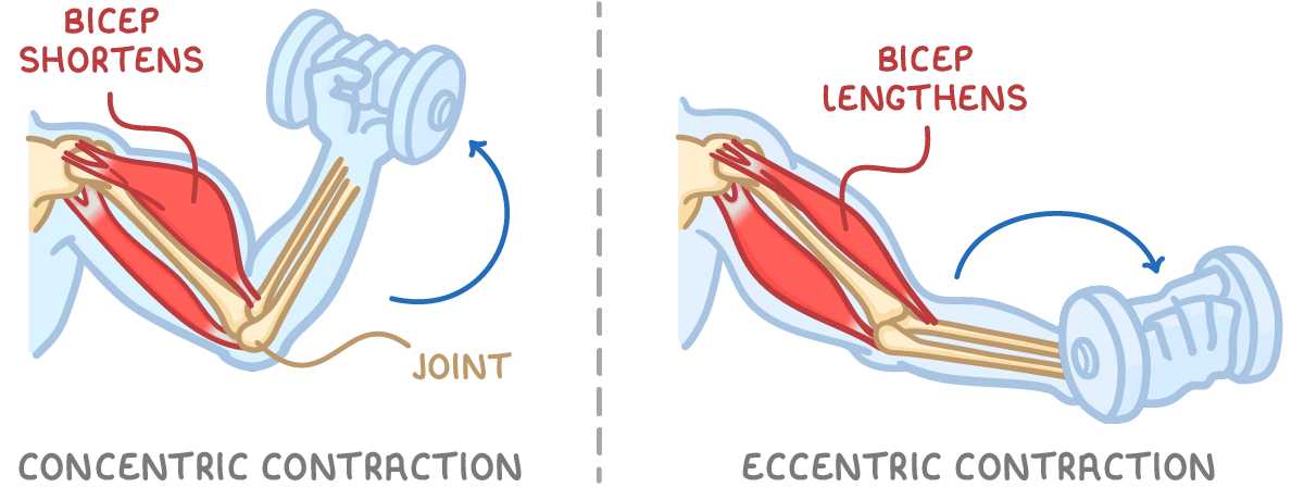
Isotonic contractions are further divided into concentric (shortening) and eccentric (lengthening) types.
Concentric contraction
A concentric contraction happens when a muscle shortens while contracting, pulling on a bone to initiate movement.
For example, during the upward phase of a biceps curl, the biceps shortens to lift the weight, flexing the elbow
Eccentric contraction
An eccentric contraction occurs when a muscle lengthens while still contracting, creating tension to control the speed of movement.
For example, during the downward phase of a biceps curl, the biceps lengthens under tension to lower the weight slowly, preventing it from dropping quickly
How antagonistic muscle pairs function
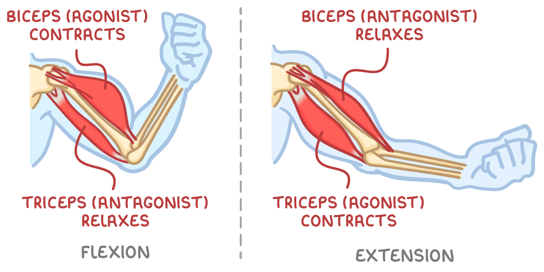
Muscles can only pull, not push, so they work in pairs called antagonistic pairs to enable movement in opposite directions at a joint. In these pairs, one muscle contracts (shortens) to pull while the other relaxes (lengthens), and they switch roles for the reverse movement. The contracting muscle is the agonist (or prime mover), responsible for the main force. The relaxing muscle is the antagonist, which opposes the agonist but must relax to allow movement. Tendons attach each muscle to two bones, but only one bone moves during the action.
Key principles of antagonistic pairs
- Opposing actions - Pairs pull in opposite directions to allow bidirectional joint movement
- Coordination - The agonist contracts while the antagonist relaxes, ensuring smooth motion without conflict
- Role reversal - When the movement direction changes, the muscles swap roles
- Tension creation - Contraction means generating tension, which can involve shortening or lengthening
Agonist and antagonist roles at key joints
Antagonistic pairs operate at various joints, with specific muscles acting as agonists or antagonists depending on the movement. The following tables outline these roles for common joints, focusing on primary muscles.
Elbow joint
| Movement | Agonist | Antagonist |
|---|---|---|
| Flexion | Biceps | Triceps |
| Extension | Triceps | Biceps |
Knee joint
| Movement | Agonist | Antagonist |
|---|---|---|
| Flexion | Hamstrings | Quadriceps |
| Extension | Quadriceps | Hamstrings |
Hip joint
| Movement | Agonist | Antagonist |
|---|---|---|
| Flexion | Hip flexors | Gluteals |
| Extension | Gluteals | Hip flexors |
Ankle joint
| Movement | Agonist | Antagonist |
|---|---|---|
| Plantar flexion | Gastrocnemius | Tibialis anterior |
| Dorsiflexion | Tibialis anterior | Gastrocnemius |
Shoulder joint
Note: The shoulder involves multiple muscles; these are the main ones for each action. The rotator cuff muscles (infraspinatus, teres minor, and subscapularis) contribute to rotation and stability.
| Movement | Agonist(s) | Antagonist(s) |
|---|---|---|
| Flexion | Front part of deltoid | Back part of deltoid |
| Extension | Back part of deltoid | Front part of deltoid |
| Rotation (outwards) | Infraspinatus, teres minor | Subscapularis |
| Rotation (inwards) | Subscapularis | Infraspinatus, teres minor |
| Adduction | Latissimus dorsi | Middle part of deltoid |
| Abduction | Middle part of deltoid | Latissimus dorsi |