1.4 - Breathing & Gas Exchange
The structure of the respiratory system
The respiratory system consists of the organs and structures involved in breathing, located primarily in the chest cavity - the space inside the chest protected by the ribcage.
Pathway of air through the respiratory system
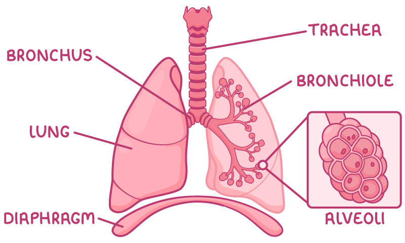
- Nose or mouth - Air enters the body through these openings.
- Trachea - Also known as the windpipe, this tube carries air towards the lungs.
- Bronchi - The trachea divides into two bronchi (singular: bronchus), with one bronchus entering each lung.
- Bronchioles - The bronchi branch into smaller tubes called bronchioles, which continue to divide and become progressively narrower.
- Alveoli - The bronchioles end in tiny air sacs called alveoli (singular: alveolus), where gas exchange occurs.
Supporting structures in the respiratory system
- Lungs - Two spongy organs that contain the bronchi, bronchioles, and alveoli, housed within the chest cavity.
- Diaphragm - A dome-shaped muscle at the base of the chest cavity.
- External intercostal muscles - Muscles located between the ribs that help expand and contract the ribcage during breathing.
- Ribcage - A bony structure that protects the lungs and heart, and moves to alter the chest cavity size.
- Capillaries - Tiny blood vessels surrounding the alveoli that facilitate gas exchange with the blood.
The mechanics of breathing in and out
Breathing, or ventilation, involves the movement of air into and out of the lungs due to changes in pressure within the chest cavity.
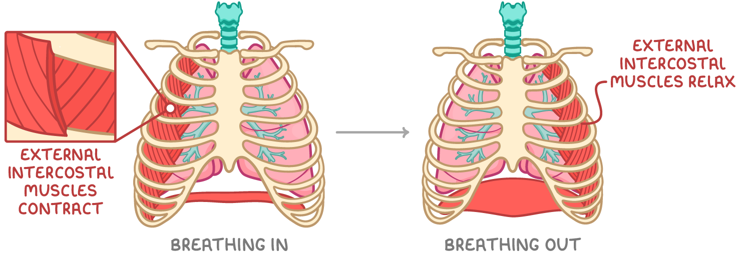
Inhalation (breathing in) is an active process
- The diaphragm contracts and flattens, moving downwards.
- The external intercostal muscles contract, pulling the ribcage upwards and outwards.
- These actions expand the chest cavity, increasing its volume.
- As a result, the air pressure inside the lungs decreases below atmospheric pressure.
- Air is drawn into the lungs through the respiratory pathway to equalise the pressure.
Exhalation (breathing out) is usually a passive process at rest
- The diaphragm relaxes and returns to its dome shape, moving upwards.
- The external intercostal muscles relax, allowing the ribcage to move downwards and inwards.
- These actions decrease the volume of the chest cavity.
- As a result, the air pressure inside the lungs increases above atmospheric pressure.
- Air is forced out of the lungs through the same pathway it entered.
Gas exchange in the alveoli through diffusion
Gas exchange occurs in the alveoli, where oxygen (O2) from inhaled air enters the blood, and carbon dioxide (CO2) from the blood is released into the air to be exhaled. This process relies on diffusion - the movement of gases from an area of higher concentration to an area of lower concentration.
Adaptations of the alveoli for efficient gas exchange
- Large surface area - Millions of alveoli provide a vast area for gases to diffuse across.
- Thin, moist walls - The alveolar walls are one cell thick and lined with moisture, allowing gases to dissolve and diffuse quickly over a short distance.
- Rich blood supply - Each alveolus is surrounded by a network of capillaries, ensuring constant blood flow for gas pickup and delivery.
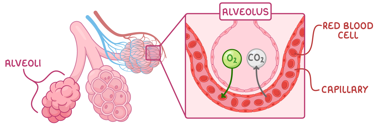
Step-by-step process of gas exchange
- Inhaled air fills the alveoli, creating a high concentration of O2 and low concentration of CO2 inside the alveoli.
- Deoxygenated blood (low in O2, high in CO2) arrives at the capillaries from the body.
- O2 diffuses from the alveoli (high concentration) into the blood (low concentration) across the thin alveolar wall.
- CO2 diffuses from the blood (high concentration) into the alveoli (low concentration).
- Oxygenated blood leaves the lungs, while CO2-rich air is exhaled.
The cardio-respiratory system and blood circulation
The cardio-respiratory system combines the cardiovascular system (heart, blood vessels, and blood) and the respiratory system to transport gases around the body.
Role of blood in gas transport
- Red blood cells - These cells contain haemoglobin, a protein that binds to O2 to form oxyhaemoglobin, allowing efficient transport.
- Oxygenated blood - Blood rich in O2 carries oxygen from the lungs to body tissues.
- Deoxygenated blood - Blood low in O2 and high in CO2 returns waste gases to the lungs.
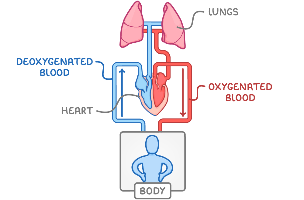
Circulation process in the cardio-respiratory system
- Oxygenated blood from the lungs returns to the heart and is pumped to the body, delivering O2 to tissues like muscles and collecting CO2.
- Deoxygenated blood returns to the heart and is pumped to the lungs.
- In the lungs, CO2 diffuses into the alveoli to be exhaled, while O2 diffuses into the blood.
- The newly oxygenated blood returns to the heart to repeat the cycle.
Definitions of key lung volumes
Lung volumes describe the amounts of air involved in breathing, which can be measured to assess respiratory function.
Main lung volumes
| Volume | Description |
|---|---|
| Tidal volume | The amount of air inhaled or exhaled in a normal breath; it increases during exercise as breaths deepen. |
| Inspiratory reserve volume (IRV) | The extra air that can be inhaled after a normal breath. |
| Expiratory reserve volume (ERV) | The extra air that can be exhaled after a normal breath. |
| Residual volume | The air remaining in the lungs after maximum exhalation. |
Spirometer traces and changes during exercise
A spirometer is a device that measures lung volumes by tracking air movement during breathing, producing a graph called a spirometer trace. This trace illustrates how breathing patterns change, particularly during exercise.
Features of a spirometer trace
- Normal breathing - Shown as small, regular waves; upward parts represent inhalation, downward parts represent exhalation.
- Maximum breaths - Larger peaks and troughs indicate breathing in and out as much as possible.
- Key measurements - The difference between a peak and dip shows tidal volume; additional differences indicate IRV, ERV, and residual volume.
Changes in breathing during exercise
During exercise, the body requires more O2 and produces more CO2.
This leads to changes in breathing:
- Increased tidal volume - Breaths become deeper, making peaks higher and dips lower on the trace.
- Increased breathing rate - More breaths per minute, shown by peaks being closer together.
- Decreased IRV and ERV - Less reserve capacity as normal breaths use more of the total lung volume.