5.7 - Using Radiation in Medicine
Medical tracers (gamma sources)
- A medical tracer is a radioactive isotope put into the body so its movement can be followed from outside.
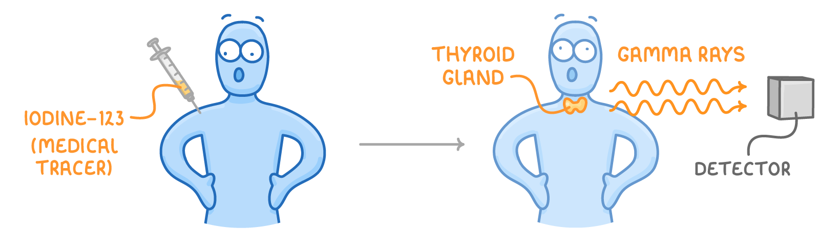
- The patient swallows it or has it injected, and an external detector tracks where the radiation comes from to show how an organ is working.
Why a gamma emitter with a short half-life
- A tracer is chosen to be a gamma emitter:
- Gamma passes out through body tissues without being absorbed, so it can be detected outside the patient.
- Gamma is only weakly ionising, so it does little damage on the way out.
- An alpha source would be useless because it would be stopped inside the body and never reach the detector. Alpha is also strongly ionising, so it would be very harmful inside the body.
- The isotope needs a short half-life, so its activity is high enough to give a clear reading at first but the radioactivity inside the patient dies away quickly. The half-life must still be long enough for the scan to be carried out.
Examples
- Iodine-123 - The thyroid gland absorbs radioactive iodine-123 the same way as non-radioactive iodine-127. The amount of gamma radiation detected from the thyroid shows whether the gland is working correctly.
- Tracer attached to a molecule - A tracer joined to a molecule taken up by cancer cells collects in those cells, so tumours show up clearly on the detector.
Radiotherapy - Treating cancer with radiation
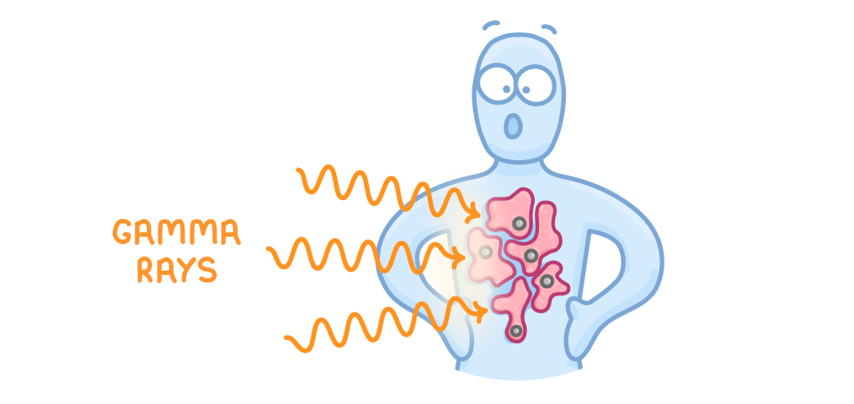
- High doses of ionising radiation kill living cells, so radiation can treat cancer by killing the cancer cells. This is called radiotherapy.
- Radiotherapy can be given from outside the body or from inside.
External radiotherapy
- Gamma rays are aimed at the tumour from outside because gamma can penetrate through the body to reach the tumour.
- The beam is carefully focused on the tumour at just the right dose, but some healthy cells near the tumour are still damaged.
- Sources used outside the body have a long half-life, so they don't need replacing often.
- The machines are kept in a shielded room to protect staff and other patients.
Internal radiotherapy
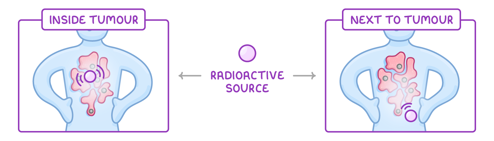
- A small amount of radioactive source is placed into or next to the tumour, as shown below.
- Alpha emitters can be injected close to the tumour. Alpha is strongly ionising, so it does a lot of damage to nearby cancer cells, while its very short range limits damage to surrounding healthy tissue.
- Beta emitters are often used in implants. Beta passes through the implant casing (which stops alpha) and has a longer range, reaching cancer cells a little further away.
- Sources used inside the body have a short half-life, to limit the time the radioactive material stays inside the patient.
Weighing the risks and benefits of medical radiation
Using radiation in medicine always means weighing the benefits against the risks.
Tracers vs radiotherapy
- Tracers - Small risk (a very small chance of causing cancer); large benefit, as tracers can diagnose serious, life-threatening conditions.
- Radiotherapy - Higher risk (causes side effects and can slightly raise the chance of another cancer later); large benefit, as it can destroy the tumour and cure the cancer.
Making the decision
- Whether to use radiation is a decision for the patient and doctor together, weighing whether the benefit is worth the risk.
- People often judge by perceived risk (how risky something feels) rather than the actual risk.
TermDefinition
TracerA radioactive substance put into something, such as a patient's body or a pipe, to show where it goes. The radiation it gives out is detected from outside.
RadiotherapyThe treatment of cancer using high doses of ionising radiation to kill cancer cells.
Perceived riskHow risky a person feels something is, which may be different from the actual risk.
Key points to remember
How were these notes?