4.8 - PET Scanning
What PET scanning is
- Positron emission tomography (PET) is an imaging technique that shows how an organ or tissue is working, rather than just its structure.
- It tracks where a radioactive tracer collects inside the body, revealing areas of high metabolic activity.
Using PET in diagnosis
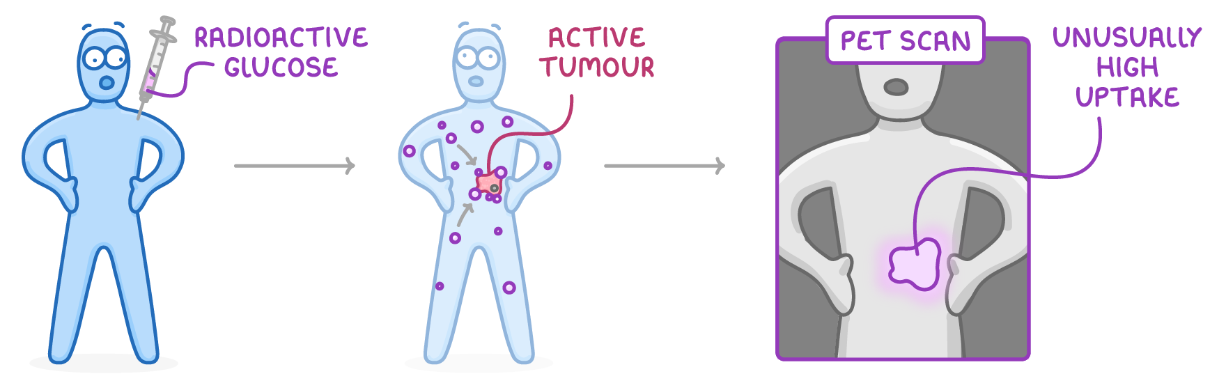
- Active cancer tumours have a much higher metabolism than healthy tissue because their cells are growing and dividing rapidly.
- The patient is given a radioactive form of glucose, which concentrates in these metabolically active cells, so the tumour shows up on the scan as an area of unusually high uptake.
The positron tracer and annihilation
- The tracer is a substance the body uses normally, such as glucose, labelled with a radioactive isotope that emits positrons.
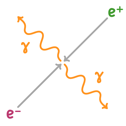
- A positron is the antiparticle of the electron: it has the same mass, but a positive charge.
- The tracer is injected into the patient and carried by the blood to the organs being studied.
Annihilation
- Inside the body, each emitted positron meets an electron.
- They annihilate: both particles are destroyed and their mass is converted into energy.
- This releases two high-energy gamma rays travelling in exactly opposite directions.
How the scanner builds an image
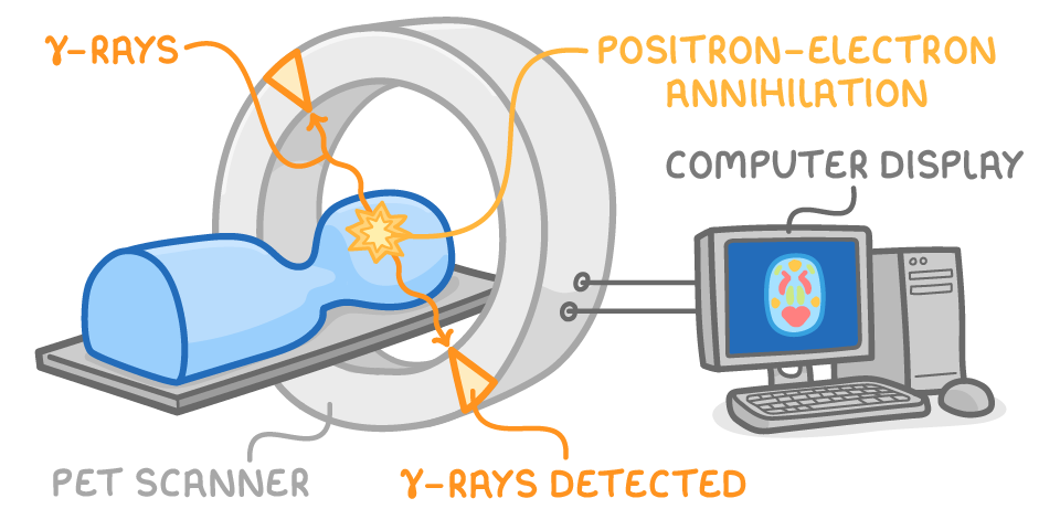
- A ring of detectors around the patient detects the gamma ray pairs.
- Because the two gamma rays travel in opposite directions, the point of emission lies on the line between the two detectors that fired.
- Combining the lines from many pairs builds up an image showing where the tracer is concentrated.
Short half-life isotopes and on-site production
- The positron-emitting isotopes used in PET have very short half-lives, which keeps the patient's radiation dose as low as possible.
- Because they decay so quickly, the isotopes must be made close to where they're used.
- Hospitals often produce them on site using a particle accelerator called a cyclotron, so their activity doesn't fall too low before the scan.
TermDefinition
Positron emission tomography (PET)An imaging technique that tracks a radioactive tracer to show how an organ or tissue is working.
PositronThe antiparticle of the electron, with the same mass but a positive charge. It is emitted from the nucleus in beta-plus decay, when a proton changes into a neutron.
AnnihilationThe process where a particle and its antiparticle meet and are destroyed, converting their mass into energy.
CyclotronA type of particle accelerator used by hospitals to produce radioactive isotopes on site.
Key points to remember
How were these notes?