6.9 - X-Rays & Gamma Rays
X-rays let us see inside the body
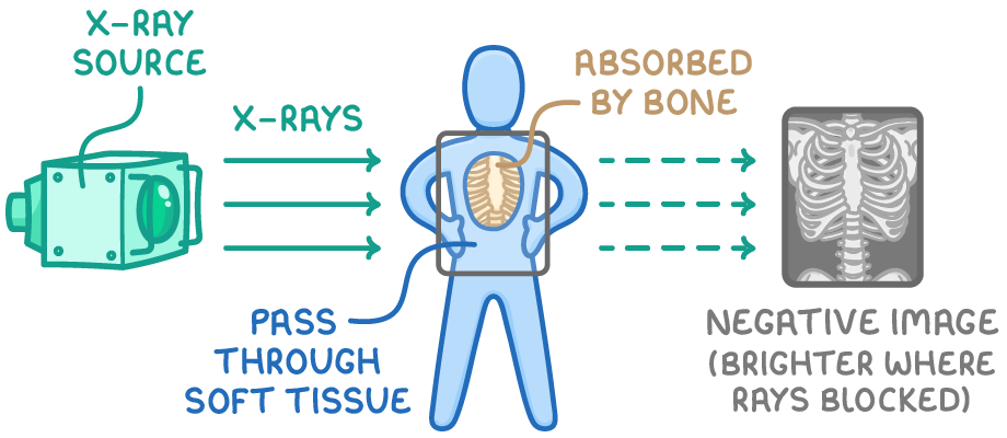
X-rays pass easily through soft tissue but are absorbed by denser material such as bone or metal.
Forming an X-ray image
- Radiation is directed through the body onto a detector plate.
- The result is a negative image: lighter areas show where X-rays were absorbed by dense material (such as bone), while darker areas show where they passed through soft tissue.
Uses of X-ray imaging
- Diagnosing broken bones and dental problems - Dense bone shows up clearly against surrounding tissue.
- CT scans - Use multiple X-rays taken from different angles to build a 3D image of the body.
- Airport security scanners - Detect hidden objects inside baggage.
Treating cancer and tracing the body
Radiotherapy
- High doses of X-rays and gamma rays kill living cells.
- In radiotherapy, beams of radiation are aimed precisely at a tumour to destroy cancer cells while minimising damage to surrounding healthy tissue.
Medical tracers
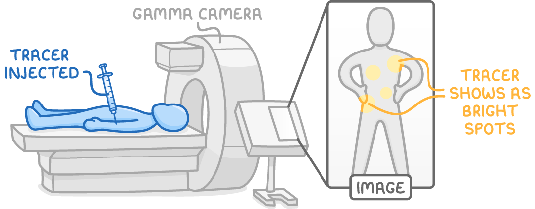
- A tracer containing a gamma-emitting source is injected or swallowed, and a gamma camera outside the body detects the emitted gamma rays to build an image.
- Gamma rays are used because they pass out through skin, soft tissue and bone without being fully absorbed.
- Tracers can be attached to molecules like glucose; cancerous tumours use more energy and take up more glucose, showing up as bright spots on the scan.
TermDefinition
CT scanA scan that uses multiple X-rays taken from different angles to build a 3D image of the body.
RadiotherapyThe treatment of cancer using high doses of ionising radiation to kill cancer cells.
TracerA radioactive substance put into something, such as a patient's body or a pipe, to show where it goes. The radiation it gives out is detected from outside.
Key points to remember
How were these notes?