5.4 - Western Front: Surgical & Medical Innovation
Key facts and dates
The Western Front during the First World War presented unprecedented medical challenges due to the scale of injuries and infections, driving rapid innovations in surgery and treatment. These advancements, born from necessity, transformed medical practice and saved countless lives both during and after the conflict.
Timeline of key events
- 1895 – Discovery of X-rays, later adapted for battlefield use.
- 1901 – Identification of blood groups by Karl Landsteiner, enabling safer transfusions.
- 1915 – Introduction of the Thomas Splint for femur fractures; Canadian doctor Lawrence Bruce Robertson begins indirect blood transfusions at casualty clearing stations.
- 1917 – Development of the Carrel-Dakin method for wound irrigation; Oswald Hope Robertson establishes the first blood bank before the Battle of Cambrai.
Innovations in treating wounds and infections
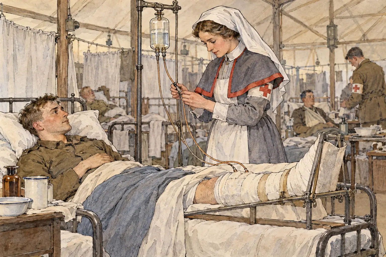
The trench warfare on the Western Front led to widespread wound infections, often caused by soil bacteria such as those responsible for tetanus and gas gangrene. Early attempts with antiseptics proved ineffective against deep, contaminated wounds, prompting surgeons to pioneer new techniques that significantly improved survival rates.
Key methods developed for wound treatment
- Wound excision (debridement) - Surgeons removed all dead, damaged, and infected tissue from wounds to prevent the spread of infection, going beyond surface cleaning to address deeper contamination.
- Carrel-Dakin method - Introduced in 1917 by Alexis Carrel and Henry Dakin, this involved continuously irrigating wounds with a sterilising solution of sodium hypochlorite, which was most effective when applied within the first day of injury and helped reduce infection in severe cases.
These approaches marked a shift towards more aggressive and systematic infection control, adapting to the unique challenges of battlefield injuries.
The introduction and impact of the Thomas Splint
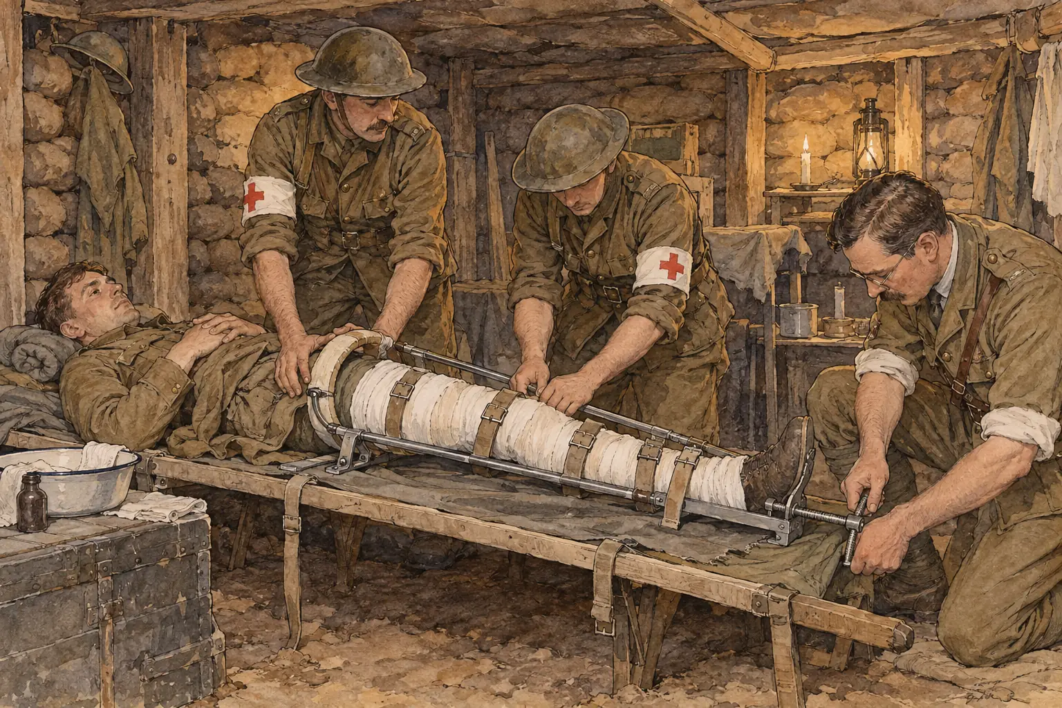
Gunshot and shrapnel wounds to the femur often resulted in compound fractures, leading to high mortality from shock and blood loss early in the war. The adoption of a pre-existing device revolutionised treatment and demonstrated how simple innovations could have profound effects.
Features and effects of the Thomas Splint
- Design and application - Invented by Hugh Thomas and adapted for wartime use from late 1915, this splint immobilised the leg by keeping it rigid and aligned during transport from the battlefield.
- Reduction in mortality - Before its widespread use, around 82% of femur fracture cases were fatal; afterwards, the rate fell to approximately 18%, primarily by minimising bleeding and shock.
- Broader significance - Medical officers at casualty clearing stations could apply the splint quickly, allowing safer evacuation of wounded soldiers and highlighting the value of stabilisation in trauma care.
Use of mobile X-ray units
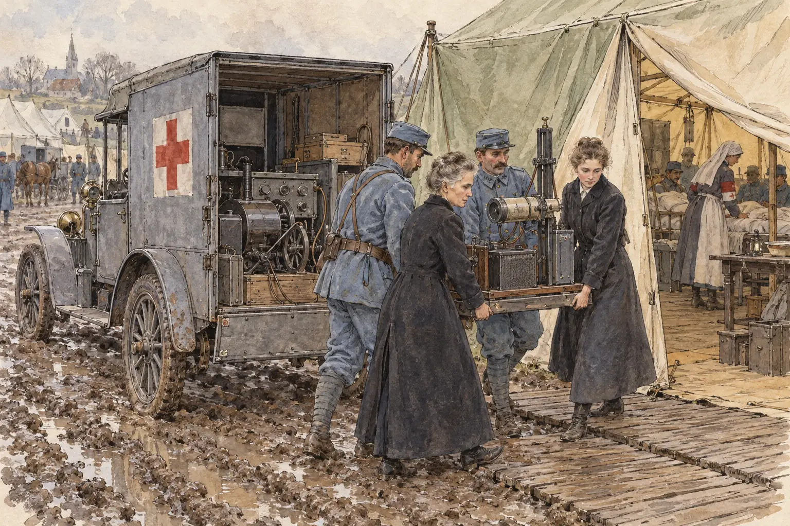
Diagnostic tools became essential for locating embedded bullets and shrapnel, which if left untreated could cause severe infections. The Western Front accelerated the practical application of X-ray technology in mobile formats to support frontline surgery.
Deployment and benefits of mobile X-rays
- Mobile units - British and French forces used X-ray vans equipped with machines and generators, positioned near casualty clearing stations to provide on-site imaging despite less sharp results than modern standards.
- Contributions from key figures - Marie Curie assisted in establishing similar units for the French army, enabling rapid identification of foreign objects and fractures.
- Impact on surgery - These units allowed surgeons to remove all fragments more accurately, reducing infection risks and improving operative success rates.
Advancements in blood transfusions and storage
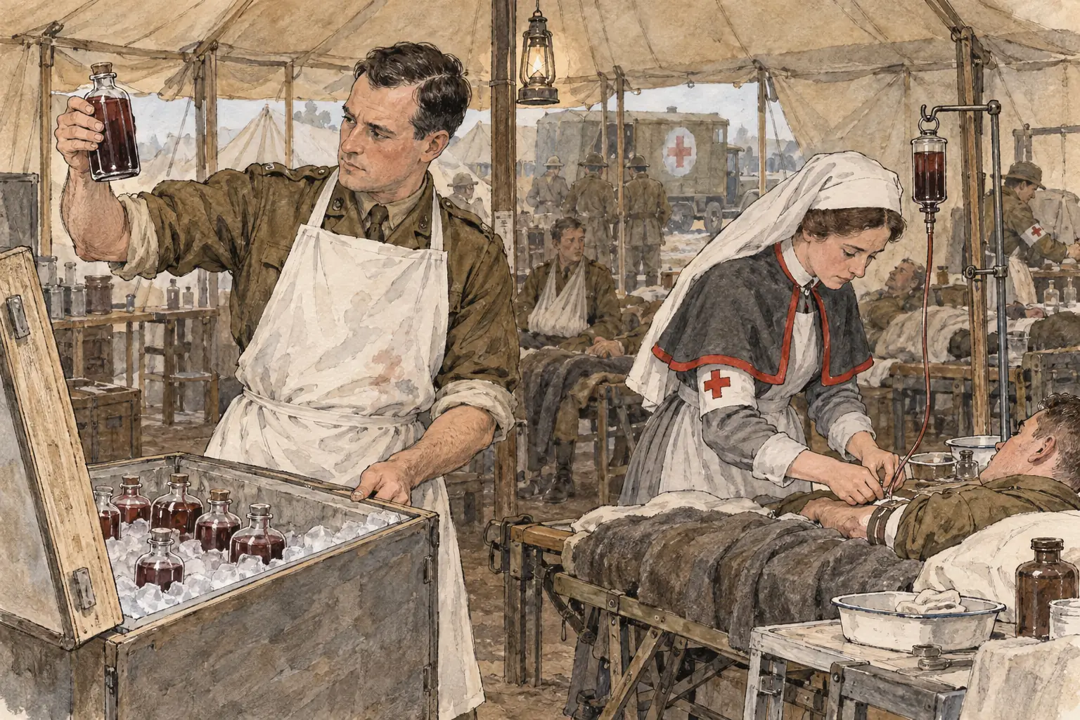
Massive blood loss from wounds frequently caused fatal shock, but the war's demands spurred progress in transfusion techniques and preservation, building on earlier discoveries like blood group compatibility.
Developments in transfusion practices
- Indirect transfusions - From 1915, doctors like Lawrence Bruce Robertson implemented transfusions at casualty clearing stations, becoming routine by 1917 among British and American forces to treat shock.
- First blood bank - In late 1917, Oswald Hope Robertson created a storage system using sodium citrate to prevent clotting, collecting 25 units of group O blood and preserving it on ice in insulated boxes for up to 28 days.
- Practical outcomes - During the Battle of Cambrai, this stored blood was used to transfuse 18 severely wounded soldiers, with 13 surviving who might otherwise have died, proving the viability of advance collection for emergencies.
These innovations laid the foundation for modern blood banking systems.
Other surgical developments accelerated by the war
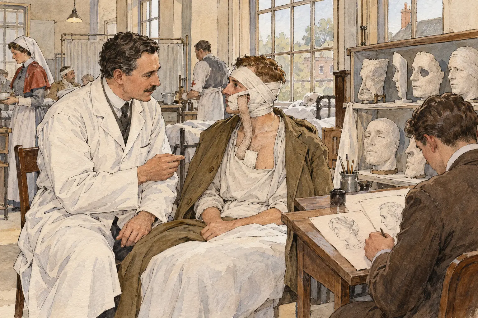
The extreme nature of injuries on the Western Front, including head trauma and facial disfigurements, forced rapid advancements in specialised fields, leading to techniques that influenced post-war medicine.
Key areas of progress
- Brain surgery - Surgeons like Geoffrey Keynes used magnets to extract metal fragments from the brain, while Harvey Cushing refined neurosurgical methods to improve outcomes in head wounds.
- Plastic surgery - Harold Gillies pioneered reconstructive procedures at Queen's Hospital in Sidcup, developing ways to rebuild faces damaged by shrapnel and burns.
- Overall legacy - The urgent need to address mass casualties encouraged experimentation, resulting in lasting improvements in trauma care, infection management, and diagnostic tools that extended beyond the battlefield.