3.2 - The Eye - Structure
The structure of the eye and its parts
- The eye is a sense organ containing receptor cells that are sensitive to light - to both its brightness (intensity) and its colour.
- The rest of the eye focuses the right amount of light onto these receptors so a clear image can form.
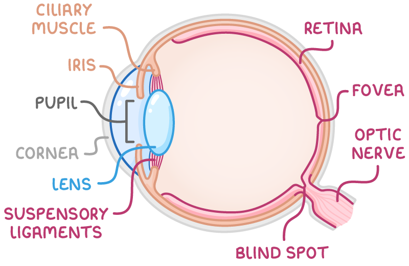
The main focusing parts
- Cornea - The transparent outer layer at the front. It refracts (bends) light as it enters and does most of the focusing.
- Iris - The coloured ring of muscle surrounding the pupil. By changing shape it controls the pupil's diameter, and so how much light gets in.
- Pupil - The hole in the middle of the iris that light passes through.
- Lens - Sits just behind the iris and pupil. It fine-tunes the focusing, bringing light to a sharp focus on the retina.
- Ciliary muscle - A ring of muscle that adjusts the lens's shape. It is attached to the lens by thread-like suspensory ligaments.
The retina and its receptor cells
- The retina lines the inside back of the eye and is the light-sensitive layer.
- It is covered in two kinds of receptor cell - rods and cones:
- Rods - Very sensitive to light, so work well in dim conditions (e.g. at night), but give only black-and-white information, no colour.
- Cones - Less sensitive, work best in brighter light, and are responsible for colour vision. There are three types of cone, each sensitive to light of a different colour.
- When light lands on these receptors, the information is turned into electrical impulses.
- The optic nerve carries these impulses from the retina to the brain, which interprets them as an image.
How rods and cones are spread across the retina
- The retina is not uniform - the two receptor types are spread differently:
- Rod cells - Mainly towards the edges (peripheral parts) of the retina, where their high sensitivity is good for seeing in dim light and detecting movement at the side of the field of view.
- Cone cells - Concentrated and packed tightly in a small central area called the fovea, which gives the sharpest, most detailed colour vision.
- This is why we turn the eye to look directly at something we want to see clearly.
Supporting and protective structures
- Sclera - The tough, white outer wall that gives the eye its shape and protects it. The cornea is the transparent part of this wall at the front.
- Conjunctiva - A thin, transparent layer covering the front surface, helping keep microorganisms out.
- Aqueous humour - Clear fluid in front of the lens.
- Vitreous humour - Clear fluid behind the lens.
- Both humours help the eye keep its shape and let light pass straight through to the retina.
- Choroid - A dark, pigmented layer between the sclera and retina. It carries blood vessels that supply the retina and absorbs stray light, stopping it reflecting around inside so the retina forms a clearer image.
- Blind spot - The small area where the optic nerve leaves the eye. It has no receptor cells, so cannot detect light. It isn't usually noticed, because the brain fills in the missing part from the surrounding information and from the other eye.
The iris (pupil) reflex
- The amount of light entering the eye must be carefully controlled:
- Too much light (very bright conditions) could damage the light-sensitive cells of the retina.
- Too little light (dim conditions) means too little reaches the retina for a clear image to form.
- The eye adjusts automatically to brightness through a reflex action that changes the size of the pupil - the hole through which light passes.
- This is an automatic reflex: light receptors detect the change in brightness and the message passes through the nervous system to the iris muscles, so the eye adjusts without any conscious effort.
The muscles of the iris
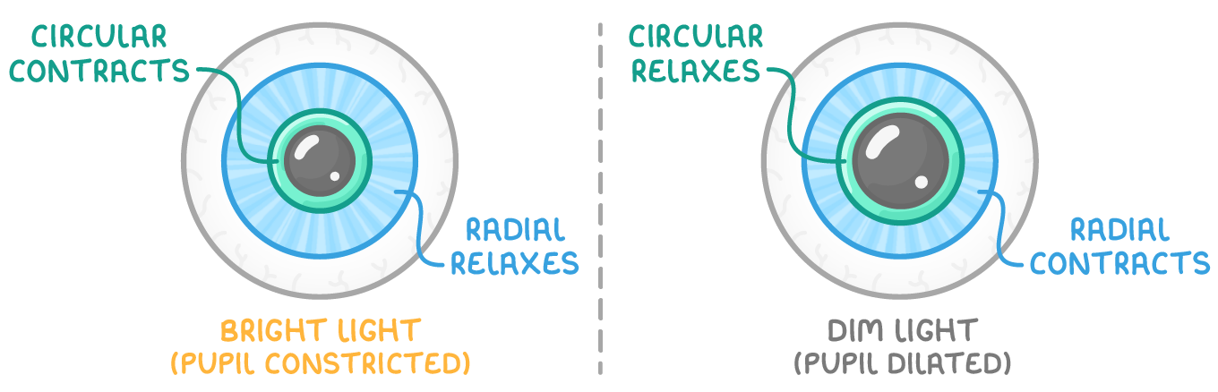
- The pupil is surrounded by the iris, which contains two sets of muscle:
- Circular muscles - Arranged in rings around the pupil.
- Radial muscles - Arranged like spokes running outwards from the pupil.
- The two sets work antagonistically - when one set contracts, the other relaxes - and between them they make the pupil smaller or larger.
How the pupil responds to light
| Conditions | Circular muscles | Radial muscles | Pupil | Effect |
|---|---|---|---|---|
| Bright light | Contract | Relax | Narrows (constricted) | Less light enters - protects the retina |
| Dim light | Relax | Contract | Widens (dilated) | More light reaches the retina |
TermDefinition
CorneaThe transparent outer layer at the front of the eye that refracts light as it enters.
IrisThe coloured ring of muscle that controls the diameter of the pupil.
PupilThe hole in the middle of the iris that light passes through.
LensThe transparent structure behind the pupil that fine-tunes the focusing of light onto the retina.
Ciliary muscleA ring of muscle that adjusts the shape of the lens.
Suspensory ligamentsThread-like fibres that attach the ciliary muscle to the lens.
RetinaThe light-sensitive layer at the back of the eye containing rod and cone receptor cells.
Optic nerveThe nerve that carries electrical impulses from the retina to the brain.
FoveaA small central area of the retina packed with cone cells that gives the sharpest colour vision.
RodsReceptor cells in the retina that are very sensitive to light but give only black-and-white information.
ConesReceptor cells in the retina that work best in brighter light and are responsible for colour vision.
Circular musclesMuscles arranged in rings around the pupil that contract to make the pupil smaller.
Radial musclesMuscles arranged like spokes around the pupil that contract to make the pupil wider.
Key points to remember
How were these notes?