2.4 - The Brain
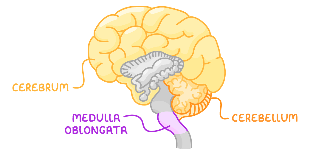
The brain's main regions
- The brain is made up of billions of interconnected neurones. It controls and coordinates everything the body does - from movement and breathing to memory and language.
- Different regions of the brain carry out different functions.
The cerebrum
- The largest part of the brain - the outer, wrinkly layer, whose surface is called the cerebral cortex.
- Divided into two halves called the cerebral hemispheres:
- The right hemisphere controls the muscles on the left side of the body.
- The left hemisphere controls the muscles on the right side.
- Responsible for the most complex behaviours: consciousness, intelligence, memory, and language.
The cerebellum
- A smaller, highly folded structure sitting underneath the cerebrum.
- Responsible for muscle coordination and balance, controlling smooth, coordinated movement.
The medulla
- Also called the medulla oblongata; it is part of the brain stem, found at the top of the spinal cord.
- Controls the unconscious activities of the body - things done automatically, without having to think about them - such as breathing and heart rate.
Investigating and treating the brain
- Mapping which part of the brain does what is difficult - the brain can't simply be opened up and watched working.
- Scientists rely on several indirect methods, and use scanners to see the brain without surgery.
Methods of investigating the brain
- Studying brain damage - If a small region is damaged, the effect on the patient shows what that region normally does. E.g. if an area at the back of the brain is damaged and the patient loses their sight, that area must be involved in vision.
- Electrical stimulation - A tiny electrode stimulates a region and the response is observed. E.g. stimulating the motor area causes muscle contraction and movement.
- Case studies - Detailed studies of individuals with abnormal brain function add further evidence.
- Examining brains after death - The brains of people who have died can be examined.
Using scanners
- CT scan - Uses X-rays to show the brain's main structures, but not their functions.
- PET scan - Uses radioactive chemicals to show which areas are active while the person is in the scanner, revealing both structure and function in real time. Highlights regions that are unusually active or inactive - useful for studying disorders such as Alzheimer's disease.
- Functional MRI (fMRI) - Shows which areas are activated while a person performs a task, such as listening to music or recalling a memory.
Why knowledge of the brain remains limited
- The brain is extremely complex and delicate.
- Some regions are hard to access.
- Results are hard to interpret - activity seen in a scanner may not match what happens in everyday life.
- Ethical limits:
- A severely brain-damaged person may be unable to give informed consent to being studied.
- Examining the brains of the dead depends on people donating them.
Why brain damage and disease are hard to treat
- Neurones in the central nervous system do not readily repair themselves, and no reliable way to repair this nervous tissue has been developed.
- Some problems sit in regions that cannot be reached safely - e.g. a tumour growing in certain parts of the brain cannot be removed surgically.
- Any treatment attempted carries the risk of causing further, permanent damage, such as the loss of a function like speech.
TermDefinition
CerebrumThe large, outer part of the brain responsible for complex behaviours such as consciousness, intelligence, memory, and language.
CerebellumA folded structure in the brain responsible for muscle coordination.
MedullaA structure in the brain that controls unconscious activities such as breathing and heart rate.
CT scanA brain scan that uses X-rays to show the brain's main structures, but not their functions.
PET scanA brain scan that uses radioactive chemicals to show which areas are active in real time.
Functional MRIA brain scan that shows which areas are activated while a person performs a task.
Key points to remember
How were these notes?