11.3 - Ventilation
The breathing-system structures
- The lungs are the organs where air is taken in and let out.
- They sit inside the thorax - the chest cavity between the neck and the diaphragm.
- The lungs are protected by the ribcage, with intercostal muscles running between the ribs.
- Below the lungs, a sheet of muscle called the diaphragm separates the thorax from the lower part of the body.
- Thin pleural membranes surround the lungs themselves.
The path of air into the lungs
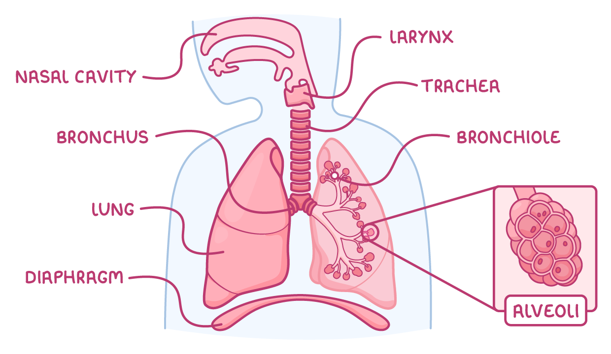
Air breathed in travels through a series of structures before reaching the sites of gas exchange:
- Nasal cavity - Air enters here first.
- Larynx - Air passes through next.
- Trachea (windpipe) - A hollow tube held open by C-shaped rings of cartilage, so it stays strong but can still move.
- Bronchi - The trachea divides into two of these tubes, one bronchus to each lung.
- Bronchioles - Each bronchus branches into progressively smaller tubes.
- Alveoli - The bronchioles end in these tiny air sacs, where gas exchange takes place.
The mechanism of breathing
- Breathing moves air into and out of the lungs by changing the volume of the thorax, and with it the air pressure inside the chest.
- Inspiration = breathing in; expiration = breathing out.
- Air always moves down the pressure difference the muscles create - from higher pressure to lower pressure.
- The ribs are moved by two sets of intercostal muscles working against each other:
- External intercostal muscles
- Internal intercostal muscles
Breathing in (inspiration)
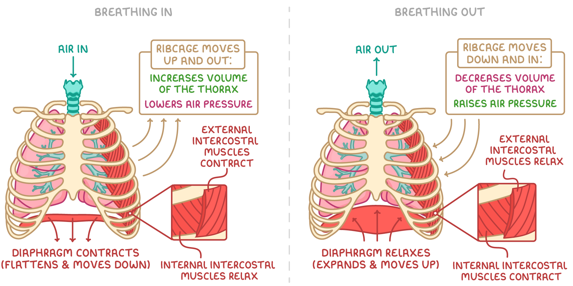
- The external intercostal muscles and the diaphragm contract; the internal intercostal muscles relax.
- The contracting external intercostals pull the ribs upwards and outwards.
- The contracting diaphragm flattens and moves down.
- Together these movements increase the volume of the thorax.
- The larger volume lowers the air pressure inside the thorax below atmospheric pressure outside.
- Air is drawn in through the trachea.
Breathing out (expiration)
- The internal intercostal muscles contract; the external intercostal muscles and the diaphragm relax.
- The ribs move downwards and inwards and the diaphragm returns to its domed shape.
- These movements decrease the volume of the thorax.
- The smaller volume raises the pressure inside the thorax above atmospheric pressure.
- Air is forced out of the lungs.
The control of breathing rate and depth
- During physical activity, the muscles respire faster to release more energy.
- This means they take in more oxygen and release more carbon dioxide into the blood.
- The brain detects the raised carbon dioxide concentration in the blood and responds by increasing both:
- Rate of breathing - More breaths each minute
- Depth of breathing - A larger volume of air with each breath
- Breathing faster and more deeply takes oxygen into the lungs and removes carbon dioxide from the blood more quickly.
- This keeps the supply of oxygen and the removal of carbon dioxide in pace with the muscles' increased demand.
TermDefinition
ThoraxThe chest cavity between the neck and the diaphragm.
DiaphragmA sheet of muscle below the lungs that separates the thorax from the lower part of the body.
TracheaThe tube, held open by rings of cartilage, that carries air from the mouth and nose to the lungs.
BronchiThe two tubes that the trachea divides into, leading to each lung.
AlveoliThe tiny air sacs in the lungs where gas exchange takes place.
InspirationThe process of breathing air into the lungs.
ExpirationThe process of breathing air out of the lungs.