10.3 - Human Reproduction & Pregnancy
The male reproductive system
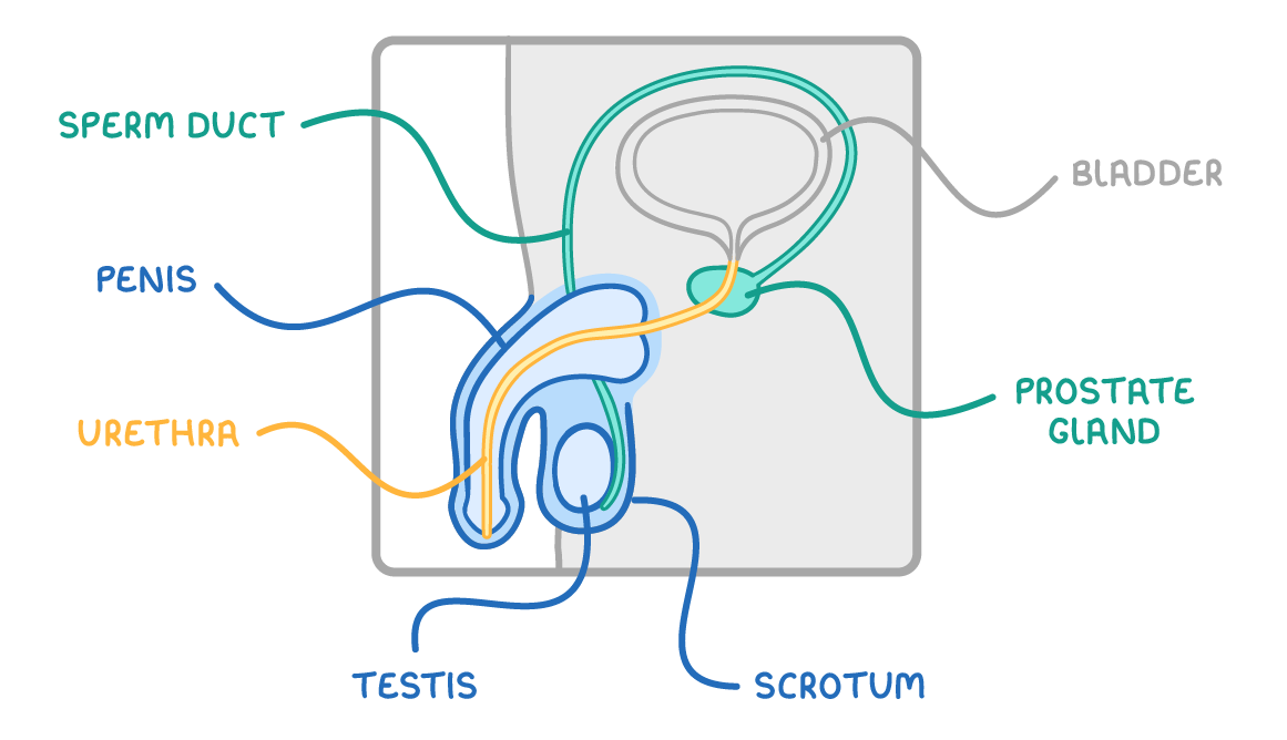
- The male reproductive system makes sperm (the male gametes) and delivers them into the female.
- It is made up of several parts, each with its own job.
Making and storing sperm
- Sperm are made in the testes.
- The two testes are held in the scrotum - a sac of skin that hangs behind the penis.
- Keeping the testes outside the main body keeps them slightly cooler than body temperature, which is the condition sperm need to be made.
Delivering the sperm
- Sperm ducts (sperm tubes) - Muscular tubes that carry sperm from the testes towards the urethra.
- Prostate gland - Adds a fluid that nourishes the sperm. The mixture of sperm and this fluid is called semen.
- Urethra - The tube running through the penis. It carries semen out of the body during ejaculation, and also carries urine out from the bladder at other times.
- Penis - Stiffens when it fills with blood, which lets it place sperm inside the female.
Female reproductive system, fertilisation, implantation
- The female reproductive system produces egg cells, holds and nourishes a developing baby, and is where fertilisation takes place.
- It is made up of several parts, each with its own job.
Parts of the female reproductive system
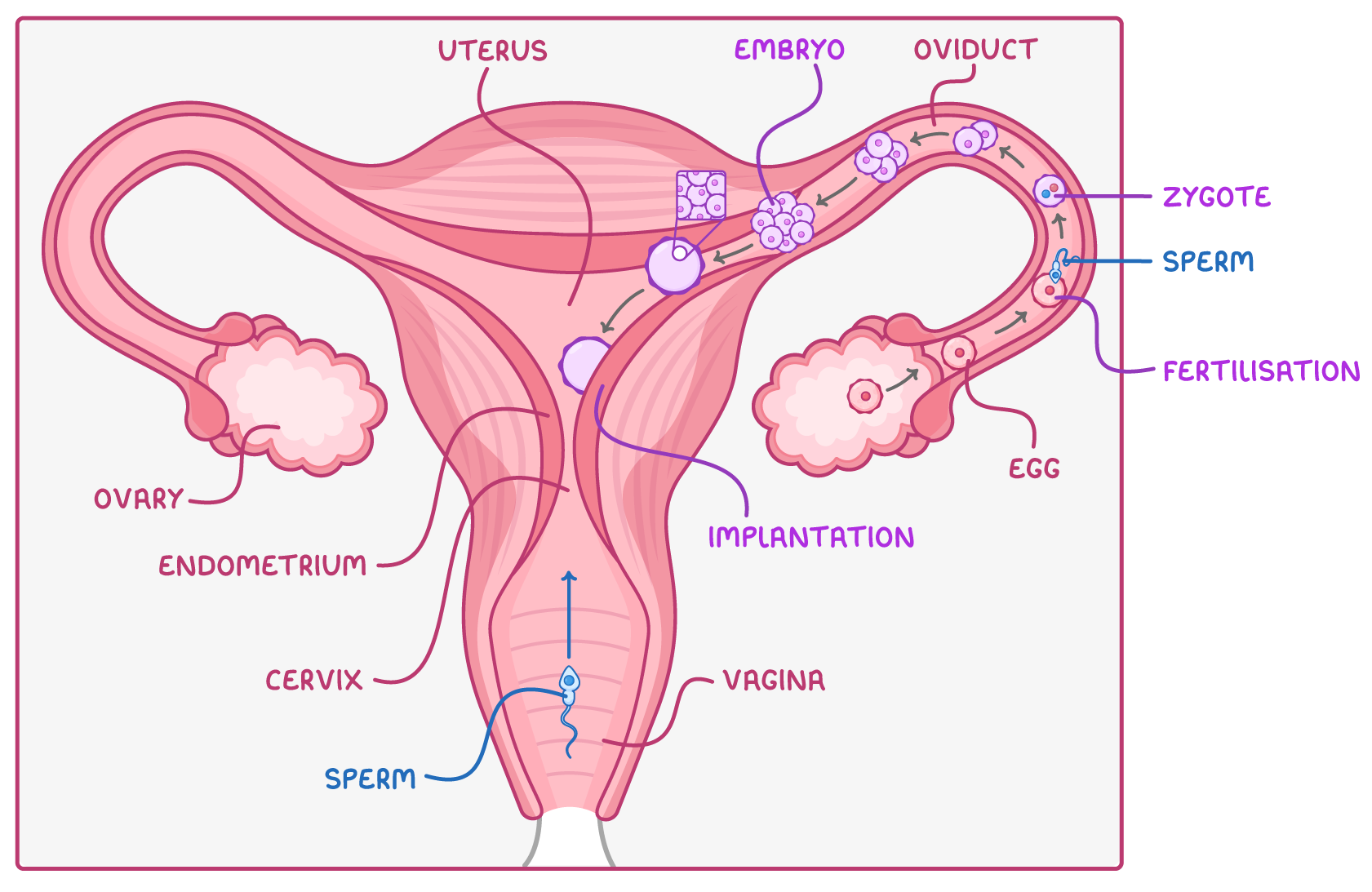
- Ovaries - Two organs that make the egg cells (ova) and also produce the sex hormones. Each month an egg is released from an ovary.
- Oviduct - A muscular tube that carries the released egg towards the uterus.
- Uterus - The organ in which an embryo can grow and develop.
- Cervix - The neck of the uterus, at its lower narrowed end; it opens into the vagina and widens during birth to let the baby pass through.
- Vagina - Where sperm are deposited; they then swim up through the cervix and uterus into the oviducts.
Fertilisation and implantation
Fertilisation happens in the oviduct.
- A sperm reaches an egg there, and the nucleus of the sperm fuses with the nucleus of the egg cell to form a single new cell called a zygote.
- Within about four days the zygote divides several times to form an embryo - a small ball of cells.
- This ball of cells travels down the oviduct to the uterus and implants into the soft, thick lining of the uterus (the endometrium), which has a good blood supply to support the implanted embryo.
This implantation marks the start of a pregnancy.
The placenta, umbilical cord and amniotic sac
- After the embryo has implanted in the lining of the uterus, several structures develop to nourish and protect it as it grows.
- At about nine weeks after fertilisation, once it has grown enough to start to look human, the developing baby is called a foetus.
The placenta - Organ of exchange
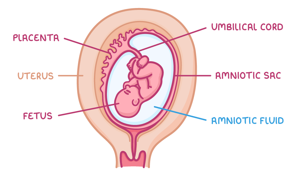
- The placenta is an organ that grows in the wall of the uterus.
- It's the organ of exchange between the mother's blood and the foetus's blood.
- It has a large surface area so substances can pass across it quickly by diffusion.
Substances moving across the placenta:
- Mother → foetus - Oxygen and dissolved nutrients (such as glucose and amino acids) supply the foetus with food and the oxygen it needs
- Foetus → mother - Waste substances, carbon dioxide and urea, pass back the other way
The umbilical cord
- The placenta is joined to the foetus by the umbilical cord.
- These substances are carried to and from the foetus in the blood vessels of the umbilical cord.
The amniotic sac and fluid
- The foetus is surrounded by a membrane called the amniotic sac, filled with a watery liquid called amniotic fluid.
- The amniotic fluid:
- Protects the foetus from knocks and bumps from outside
- Supports its weight as it grows
- Lets it move around
TermDefinition
Semen
Zygote
Embryo
Foetus
Placenta
Amniotic fluid