7.1 - Using a Light Microscope
The technique
Aim
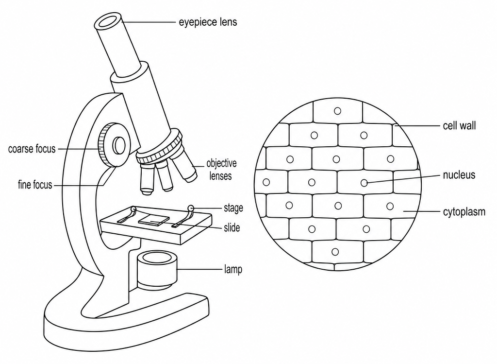
To prepare a slide of cells (for example onion epidermis), observe them under a light microscope, make a labelled drawing, and calculate the magnification or the real size of a cell.
Background
- Cells are far too small to see with the naked eye.
- A light microscope magnifies them with two lenses, the eyepiece lens and the objective lens. The total magnification is the eyepiece lens magnification multiplied by the objective lens magnification, so a ×10 eyepiece with a ×40 objective gives ×400.
- Magnification is how many times bigger the image is than the real object.
- A cell is too small to measure with a ruler on its own, but under the microscope you can count how many cells fit across a width you have measured with a clear ruler on the stage. In a photograph of known magnification, you measure the image instead and divide by the magnification.
- So in this practical you prepare a stained slide of onion cells, make a labelled drawing, and measure a cell, so you can work out its real size.
Method
- Prepare the onion layer - Peel a thin, single layer of epidermis from an onion and lay it flat on a clean glass slide.
- Stain the cells - Add a drop of iodine solution.
- Cover the slide - Lower a coverslip slowly at an angle, using a mounted needle.
- Focus on low power - Put the slide on the stage and start with the lowest-power objective lens. Focus with the coarse focus first, then sharpen the image with the fine focus.
- Switch to higher power - Turn to a higher-power objective lens and refocus to see the cells in more detail.
- Draw and measure - Make a large, labelled drawing of a few cells. To measure them, put a clear ruler on the stage under a low-power objective to find the width of the field of view, then count how many cells fit across it and divide the width by that number.
Safety
- Iodine solution - It's an irritant, so wear eye protection, use a dropper so it doesn't spill, and wash any off your skin at once.
- Slides and coverslips - They break easily into sharp pieces, so handle them by the edges and sweep up any breakages with a brush, not your fingers.
- Mounted needle - Keep it pointed away from you.
Worked example - Real width of a cell
A photograph of a plant cell was taken at a magnification of ×1,500. On the photograph the cell measures 60 mm across. Find the real width of the cell in micrometres.
Step 1 - Rearrange the formula for the actual size
Magnification = image size ÷ actual size, so actual size = image size ÷ magnification.
Step 2 - Convert millimetres to micrometres
There are 1,000 µm in 1 mm.
0.04 × 1,000 = 40 µm
The real width of the cell is 40 µm.
Step 3 - Check the answer the other way round
A cell 0.04 mm wide that is shown 60 mm wide has been magnified this many times.
60 ÷ 0.04 = 1,500
This matches the stated magnification of ×1,500. Magnification has no units, because it's simply how many times bigger the image is.
Common exam questions
Question 1
Why is a stain such as iodine added to the slide?
Most cell parts are colourless, so the stain makes them show up under the microscope.
Question 2
Why is the coverslip lowered slowly at an angle?
To stop air bubbles being trapped underneath, which would block the view of the cells.
Question 3
Why do you start with the lowest-power objective lens?
It gives the widest view, so the cells are easier to find before you zoom in.
Question 4
What should a good biological drawing include?
Clear, unbroken lines with no shading, drawn large, with labels on straight ruled lines and the magnification stated.