2.3 - The Digestive System
The organs of the digestive system
- The digestive system breaks food down into small molecules the body can absorb and use, and passes out what's left.
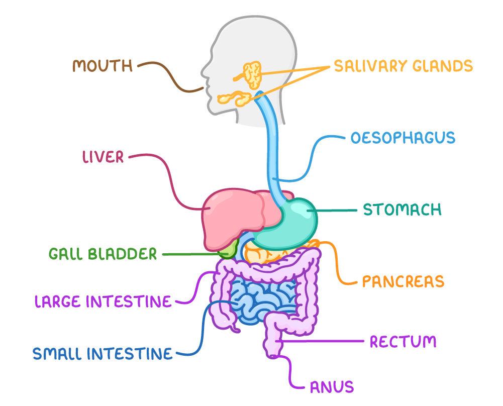
- Its central part is the alimentary canal (the gut) - a long muscular tube running from mouth to anus.
- The alimentary canal plus the organs that empty into it make up the whole digestive system.
- Food travels along the canal in one direction, and each region has its own job.
The alimentary canal, from mouth to anus
- Mouth - Takes food in and starts breaking it down. The teeth break food into smaller pieces, and the salivary glands release saliva, which contains a digestive enzyme that starts to digest starch.
- Oesophagus - The muscular tube carrying food from the mouth down to the stomach.
- Stomach - A muscular bag that churns food with its muscular walls into a paste. It releases gastric juice containing hydrochloric acid, which kills many bacteria taken in with food and makes the contents strongly acidic - the condition the stomach's own enzyme needs.
- Small intestine - A long, narrow tube whose first part is the duodenum and last part is the ileum. Digestion is completed here, and the small, soluble products of digestion (plus most of the water) are absorbed through the wall into the blood.
- Large intestine (the colon) - Most of the remaining water is absorbed back into the body, leaving a semi-solid waste.
- The waste, called faeces, is stored in the rectum (the last part of the large intestine) before being passed out through the anus.
Three organs sit alongside the canal and release their secretions into it:
- Liver - Makes bile, a fluid that helps with the digestion of fats.
- Gall bladder - Stores the bile made by the liver and releases it, through the bile duct, into the small intestine.
- Pancreas - Makes digestive enzymes and releases them into the small intestine to help complete digestion.
The villus and its adaptations
- The small intestine is adapted to absorb the products of digestion quickly and efficiently.
- It is very long, giving plenty of time to digest and absorb food before what's left reaches the end.
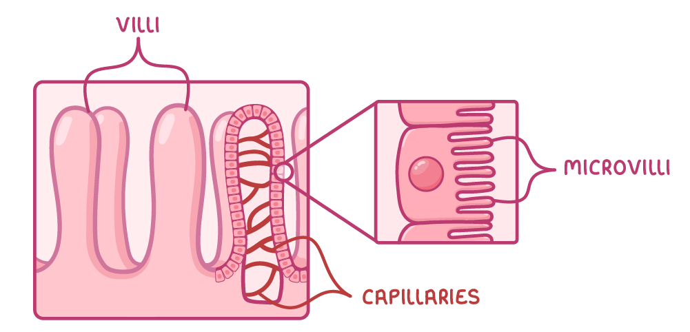
- Its inner wall has an enormous surface area - it is folded and covered in millions of tiny finger-like projections called villi.
- Each villus is a microscopic finger-like extension sticking out into the inside of the gut.
- The surface cells of each villus are themselves covered in even smaller projections called microvilli.
- Together, the length, folds, villi, and microvilli give a very large surface area, so a great deal of food can be absorbed at once.
- Most of this absorption takes place in the ileum, the last and longest part of the small intestine, whose inner wall is covered in villi.
A short diffusion distance
- A villus has a single, thin layer of surface cells - the wall is only one cell thick, and the cells have thin, permeable membranes.
- So the products of digestion have only a very short distance to travel from the gut into the blood, making absorption fast.
A rich blood supply
- Inside each villus is a network of blood capillaries lying very close to the thin surface.
- The capillaries absorb the soluble products - such as glucose and amino acids - and carry them away in the blood.
- This constant removal keeps the concentration inside the villus lower than in the gut, maintaining a steep concentration gradient that drives more products to diffuse in.
- Some absorption also uses active transport, which is why the surface cells contain many mitochondria to provide the energy needed.
TermDefinition
Alimentary canalThe long muscular tube that food passes along, from the mouth to the anus.
VillusA microscopic finger-like extension sticking out into the inside of the gut.
Key points to remember
How were these notes?