1.4 - The Brain
The brain: an overview of its major structures
The brain is the control center of the body, orchestrating everything from basic survival functions to complex thoughts and emotions. Each part of the brain has specialized roles, and together, they shape behavior and mental processes. Understanding these structures helps explain how we think, feel, and act.
Key brain regions and their general functions
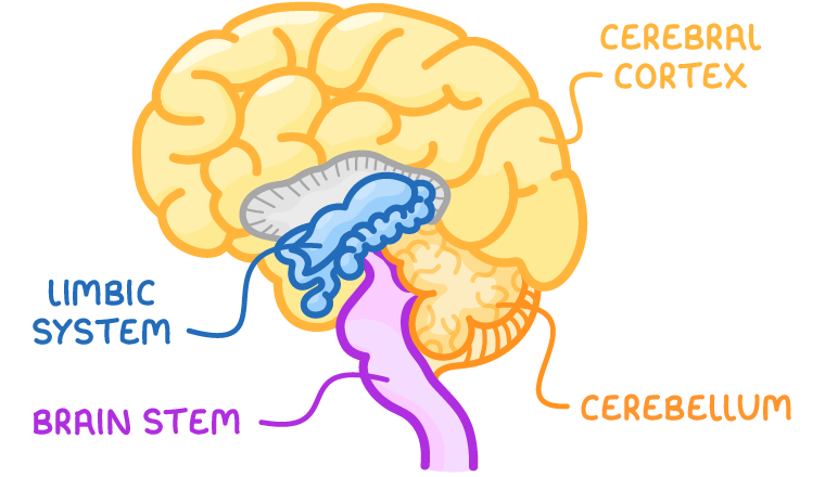
- Brain stem - Located at the base of the brain, it handles fundamental life-sustaining functions.
- Cerebellum - Positioned at the back of the brain, it manages movement and balance.
- Cerebral cortex - The outer layer of the brain, divided into two hemispheres, it governs higher cognitive processes.
- Limbic system - A set of structures deep within the brain, it regulates emotions and memory.
Each of these regions interacts to ensure the body and mind function harmoniously. Let's dive deeper into their specific contributions.
The brain stem: controlling basic survival functions
The brain stem is a critical structure connecting the brain to the spinal cord. It oversees involuntary processes that keep us alive, often without conscious awareness.
Key components and functions of the brain stem
- Medulla - Regulates vital functions such as breathing, heart rate, and blood pressure. This ensures oxygen delivery and circulation even when we're asleep.
- Pons - Acts as a bridge for signals between different brain regions and assists with functions like sleep and arousal.
These automatic processes are essential for survival, forming the foundation upon which other brain activities build.
The reticular activating system (RAS)
The reticular activating system is found within the brain stem and influences alertness and attention. It plays a crucial role in regulating arousal, wakefulness, and consciousness, helping to filter incoming sensory information and determine what requires our attention.
This system helps us stay awake and focused, playing a subtle but significant role in daily activities.
The cerebellum: coordinating movement and balance
Located at the base of the brain, just above the brain stem, the cerebellum (often called the "little brain") is crucial for physical coordination. Its intricate folds increase surface area, allowing precise control over motor skills.
Functions of the cerebellum
- Muscle coordination - Smooths out voluntary movements, ensuring actions like writing or walking are fluid rather than jerky.
- Balance - Maintains posture and equilibrium, preventing falls by adjusting body position.
- Procedural learning - Helps store learned motor skills, such as riding a bike, so they become automatic over time.
Damage to the cerebellum can lead to clumsy movements or difficulty maintaining balance, highlighting its importance in everyday tasks.
The cerebral cortex: the center of higher thought
The cerebral cortex is the wrinkled outer layer of the brain, divided into two hemispheres (left and right) connected by a thick band of nerve fibers called the corpus callosum. This structure is responsible for complex mental processes and is further divided into four lobes, each with specialized functions.
Structure of the cerebral cortex
- Hemispheres - The brain is split into left and right halves, each controlling the opposite side of the body and specializing in certain tasks.
- Corpus callosum - A bridge of nerve fibers allowing communication between hemispheres, ensuring coordinated processing.
- Lobes - Four distinct regions (frontal, parietal, temporal, and occipital) handle different sensory and cognitive functions.
This organization allows the brain to process vast amounts of information efficiently by dividing tasks among specialized areas.
Functions of the four lobes of the cerebral cortex
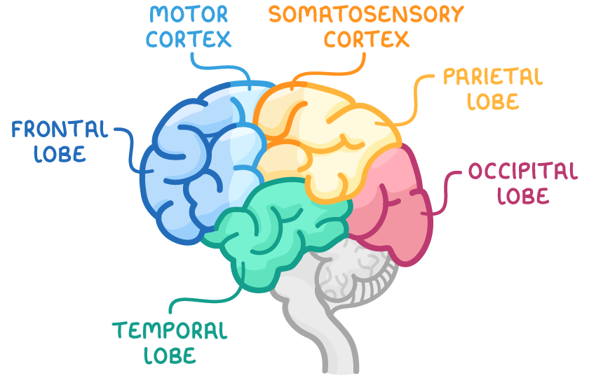
Frontal lobes:
- Location - Just behind the forehead.
- Primary functions - Govern complex cognitive processes, higher-order thinking, and executive functioning (planning, decision-making, problem-solving) via the prefrontal cortex.
- Motor cortex - Found at the rear of the frontal lobes, controls voluntary skeletal movements like waving or kicking.
Parietal lobes:
- Location - Near the back crown of the brain.
- Primary functions - Contain association areas that integrate and organize information from various senses.
- Somatosensory cortex - Processes touch, pressure, temperature, and pain sensations, mapping these inputs to specific body parts.
Temporal lobes:
- Location - On the sides of the brain, near the ears.
- Primary functions - Handle auditory processing (sound) and linguistic processing (language comprehension and memory for words).
Occipital lobes:
- Location - At the rear of the brain.
- Primary functions - Process visual information, interpreting shapes, colors, and motion from the eyes.
These lobes work together, allowing us to perceive the world, think critically, and respond appropriately to our environment.
The limbic system: regulating emotion and memory
Nestled deep within the cerebral cortex, the limbic system is a group of interconnected structures that play a central role in emotions, motivation, and memory formation. It acts as a bridge between basic instincts and higher thought processes.
Key components of the limbic system
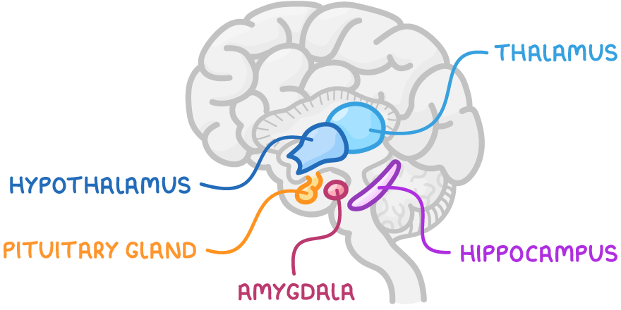
- Thalamus - Acts as a sensory relay station, directing incoming sensory information (except smell) to appropriate cortical areas for processing.
- Hypothalamus - Regulates basic drives like hunger, thirst, and body temperature, and controls the pituitary gland to release hormones affecting stress and growth.
- Pituitary gland - Known as the "master gland," it releases hormones influencing other endocrine glands, impacting metabolism, reproduction, and stress responses.
- Hippocampus - Critical for forming new memories and spatial navigation, converting short-term memories into long-term storage.
- Amygdala - Processes emotions, especially fear and anger, and triggers the fight-or-flight response during perceived threats.
This system links emotional experiences with memory, explaining why strong emotions often create lasting recollections.
Hemispheric specialization and split-brain research
The brain's two hemispheres are not identical in function; they often specialize in different tasks. Research on split-brain patients has provided deep insights into this specialization.
Understanding hemispheric specialization
- Left hemisphere - Typically dominates language processing, logical thinking, and analytical tasks.
- Right hemisphere - Excels in spatial reasoning, face recognition, and emotional tone interpretation.
This division of labor allows the brain to handle diverse tasks efficiently, though both hemispheres collaborate for most activities through the corpus callosum.
Split-brain research
Split-brain research involves studying patients whose corpus callosum has been severed, often as a treatment for severe epilepsy (a neurological disorder characterized by recurrent seizures). This surgery isolates the hemispheres, revealing their unique roles.
Key findings from split-brain studies
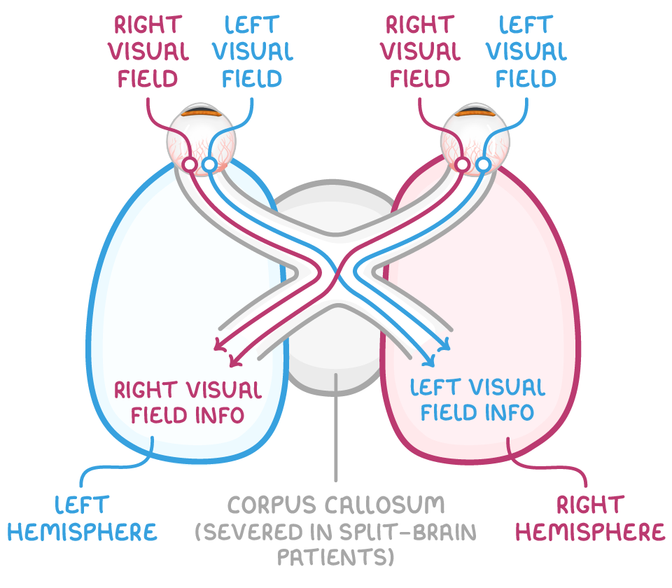
- Contralateral organization - Each hemisphere controls the opposite side of the body; for example, the right hemisphere processes input from the left visual field.
- Testing specialization - Researchers present information to one visual field at a time to isolate which hemisphere processes it, showing distinct strengths.
- Language centers - Language functions, such as speech production and comprehension, are usually in the left hemisphere.
Language-specific areas
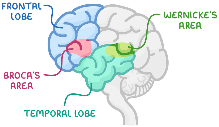
- Broca's area - Located in the left frontal lobe, responsible for speech production. Damage here can cause Broca's aphasia, where individuals struggle to form words but can understand language.
- Wernicke's area - Found in the left temporal lobe, handles speech comprehension. Damage leads to Wernicke's aphasia, where speech is fluent but nonsensical, and comprehension is impaired.
These findings highlight how specific brain regions underpin critical functions like communication.
Brain plasticity: adapting to change
Brain plasticity, also known as neuroplasticity, refers to the brain's remarkable ability to rewire itself by forming new connections or modifying existing ones. This adaptability is vital for learning and recovery from injury.
How brain plasticity works
- Throughout development - Plasticity is most pronounced in childhood, as the brain grows and learns, but it continues into adulthood.
- Response to damage - If one part of the brain is injured, other areas can sometimes take over its functions, compensating for the loss.
- Learning and experience - Repeated activities strengthen neural pathways, making skills or memories more automatic over time.
This flexibility underscores the brain's resilience, allowing recovery from trauma or adaptation to new challenges.
Research methods for studying the brain
Scientists use various techniques to explore how the brain's structures and functions relate to behavior and mental processes. These methods provide windows into the brain's inner workings, often balancing precision with ethical considerations.
Key research methods
- Electroencephalogram (EEG) - Measures electrical activity in the brain via electrodes on the scalp, useful for studying sleep patterns or seizure activity.
- Functional Magnetic Resonance Imaging (fMRI) - Detects blood flow changes in the brain, indicating active areas during specific tasks like thinking or feeling emotions.
- Case studies - In-depth analyses of individuals with unique brain conditions or injuries, offering insights into localized brain functions.
- Surgical procedures (lesions) - Involve studying the effects of intentional or accidental damage to specific brain areas, often in animals, to understand regional roles.
Each method contributes uniquely to our understanding, though ethical concerns, especially with invasive techniques, must always be evaluated to protect participants.