2.1 - Localisation & Lateralisation of Brain Function
The concept of localisation of function in the brain
Localisation of function refers to the idea that specific areas of the brain are responsible for particular mental processes and behaviours. Rather than the brain working as a single, uniform entity, different regions specialise in tasks such as vision, language, or movement.
Key functional areas of the cerebral cortex
The cerebral cortex, the outer layer of the brain, is divided into regions that handle specific functions.
Major functional areas and their roles
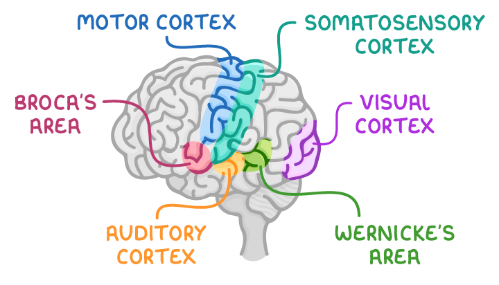
- Motor cortex - Controls voluntary movements.
- Somatosensory cortex - Processes sensory input related to touch, pain, temperature, and proprioception (awareness of body position).
- Broca's area - Manages the production of speech.
- Visual cortex - Interprets visual information received from the eyes.
- Auditory cortex - Handles sound processing from the ears.
- Wernicke's area - Responsible for language comprehension.
Hemispheric lateralisation and the role of the corpus callosum
The brain is divided into two halves, known as the left and right hemispheres, each with dominant roles in certain functions. These hemispheres are connected by a bundle of nerve fibres called the corpus callosum, which facilitates communication between them.
Differences between the hemispheres
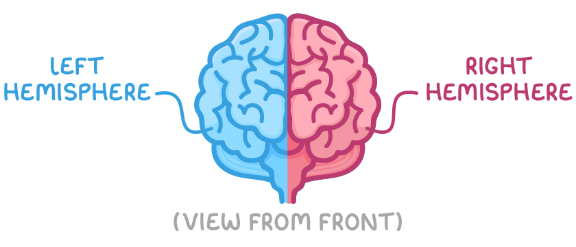
- Left hemisphere - Typically dominates language functions, with Broca's and Wernicke's areas usually located here in most individuals. It also governs logic, analytical thinking, and problem-solving skills.
- Right hemisphere - Focuses on spatial awareness, emotional processing, and recognition of faces.
- Hemispheric lateralisation - Refers to the specialisation of functions in each hemisphere.
Function of the corpus callosum
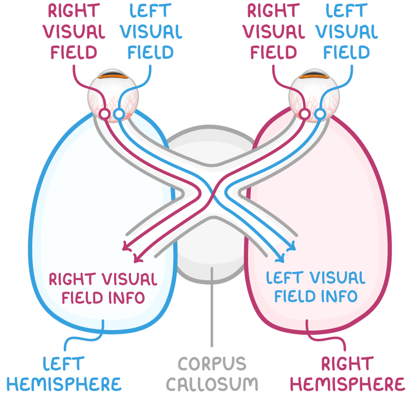
- Cross-hemisphere communication - Allows the left and right hemispheres to share information.
- Opposite-side processing - Each hemisphere primarily deals with input from the opposite side of the body. For instance, the right visual field is processed by the left hemisphere, and vice versa.
- Integration of information - Information received by one hemisphere can be transferred via the corpus callosum to the other side for further processing or response.
Split brain research and Sperry's (1968) study
In severe cases of epilepsy, a surgical procedure to sever the corpus callosum is sometimes performed to prevent the spread of seizures across the brain.
Overview of split brain surgery
- Purpose of surgery - Cutting the corpus callosum stops epileptic seizures from spreading between hemispheres.
- Side effect - Prevents information transfer between hemispheres.
- Research opportunity - Split brain patients provide insights into hemispheric specialisation.
Sperry's (1968) study on split brain patients
Method:
- Involved 11 participants who had undergone split brain surgery for uncontrollable epilepsy, alongside a control group with intact corpus callosum.
- In one experiment, participants covered one eye and fixated on a central point on a screen.
- Images were briefly flashed to either the left or right side of the screen, too quickly for eye movement to adjust.
Results:
- When images appeared in the right visual field, both split brain and control participants could verbally describe or write about them.
- However, when images were shown in the left visual field, split brain participants could not name or write about what they saw but could select a matching object with their left hand, despite not knowing why they chose it.
Conclusions:
- Demonstrates that the left hemisphere, receiving input from the right visual field, handles language production, converting visual data into words.
- The right hemisphere, receiving input from the left visual field, cannot produce language but can generate non-verbal responses.
- Normally, the corpus callosum allows information to cross for processing, but in split brain patients, this integration is absent.
Evaluation:
- The combination of case studies and experiments provided both detailed qualitative insights and measurable quantitative data, enhancing reliability and validity.
- However, the small sample size of 11 limits generalisation to the wider population, and finding more split brain patients for study is challenging.
- Additionally, pre-existing brain damage from epilepsy and medication use may have influenced results, making it unclear if findings apply to non-epileptic individuals.
- The artificial experimental setup also raises concerns about ecological validity, as it may not reflect real-world scenarios.
Evaluation of research into localisation of function
Strengths of localisation research
- Supporting evidence - Studies like Sperry's align with observations from brain injury cases, where damage to specific areas results in predictable deficits, reinforcing the concept of localisation.
- Technological advances - Modern brain imaging techniques allow precise mapping of brain activity, confirming the roles of specific regions during tasks.
- Practical applications - Understanding localisation aids in diagnosing and treating neurological conditions by targeting specific brain areas for intervention.
Limitations of localisation research
- Over-simplification - While certain functions are localised, many complex processes involve networks across multiple brain regions, suggesting a more distributed model.
- Individual variability - Brain organisation can differ between individuals, with some showing atypical lateralisation.
- Ethical and practical constraints - Research often relies on rare cases like split brain patients, limiting sample sizes and the ability to draw broad conclusions. Additionally, experimental settings may not reflect natural brain functioning.