2.2 - Localisation & Lateralisation of Brain Function
Localisation of function and key brain areas
Localisation of function refers to the idea that specific regions of the brain are responsible for particular tasks.
The functions of key brain areas
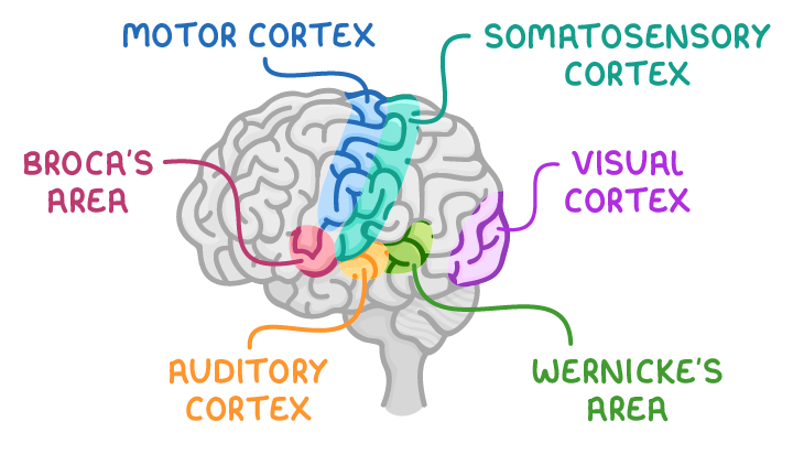
| Brain area | Primary function |
|---|---|
| Motor cortex | Controls voluntary movements. |
| Somatosensory cortex | Processes sensory information related to touch, pain, temperature, and proprioception (awareness of body position). |
| Broca's area | Handles the production of speech. |
| Wernicke's area | Involved in understanding language. |
| Visual cortex | Processes visual information received from the eyes. |
| Auditory cortex | Processes sounds and auditory information from the ears. |
Brain hemispheres and hemispheric lateralisation
The brain is divided into two halves, known as the left hemisphere and the right hemisphere, which are connected by the corpus callosum. Each hemisphere tends to dominate certain functions, a phenomenon known as hemispheric lateralisation.
Functions associated with each hemisphere
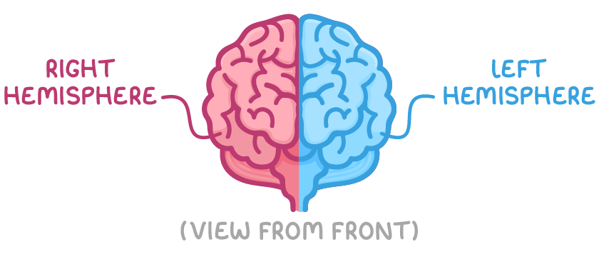
- Left hemisphere:
- Primarily manages language-related tasks, including those handled by Broca's and Wernicke's areas (which are typically located only in this hemisphere for most people).
- Focuses on logical thinking, analytical skills, and problem-solving abilities.
- Right hemisphere:
- Specialises in spatial awareness.
- Deals with emotional processing and recognising faces.
Communication between brain hemispheres
The hemispheres work together by exchanging information.
How hemispheres process and share information:
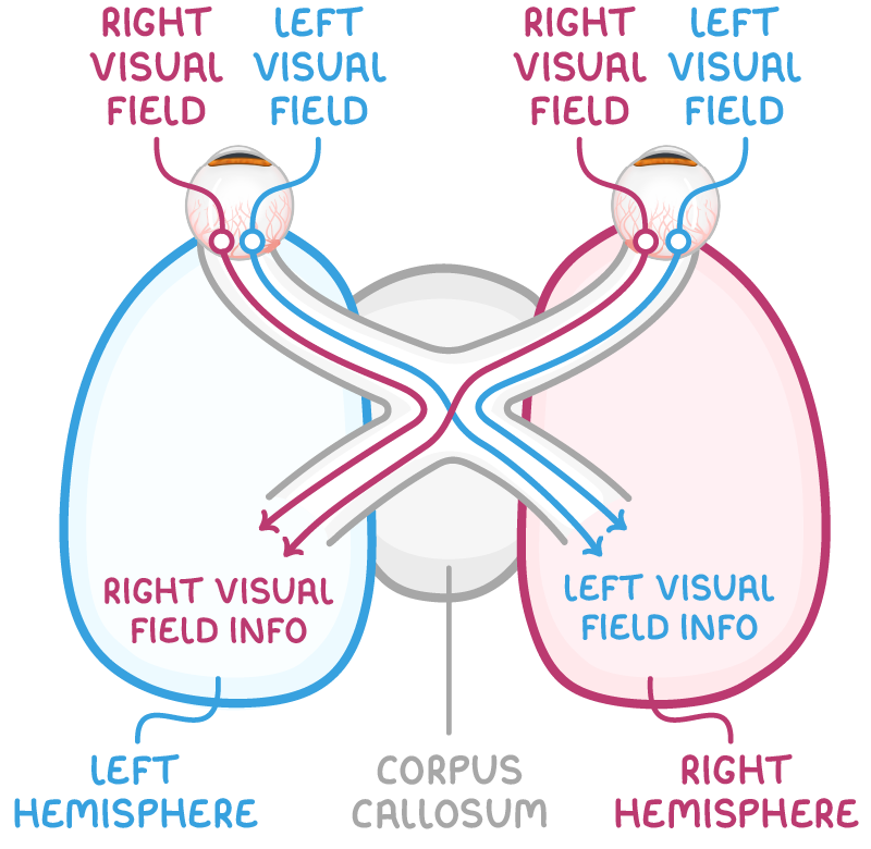
- Each hemisphere receives and processes input from the opposite side of the body.
- The corpus callosum acts as a bridge, allowing data to transfer between hemispheres.
- For example, visual information from the right side of the visual field travels to the left hemisphere's visual cortex, while left-side information goes to the right hemisphere's visual cortex; if needed, it crosses via the corpus callosum for further processing.
Split brain surgery and Sperry's research
Split brain surgery involves cutting the corpus callosum to prevent severe epileptic seizures from spreading between hemispheres.
Effects of split brain surgery:
- Severing the corpus callosum stops information transfer, isolating the hemispheres.
- This allows researchers to study each hemisphere's independent functions, as data cannot cross over.
- Patients with this surgery have been key in experiments exploring brain specialisation.
Sperry (1968) study on split brain effects
Sperry investigated how disconnecting the hemispheres affects perception and response, using patients who had undergone the surgery.
Method
The research combined case studies with experiments on 11 participants who had split brain surgery for uncontrollable epilepsy. A control group with intact hemispheres was included for comparison. In a key experiment, participants covered one eye and fixated on a central point while images were briefly flashed to either the left or right side of a screen.
Results
When images appeared in the right visual field, all participants (including split brain patients) could verbally describe or write about them. However, split brain participants could not name or write about images in the left visual field, though they could select matching objects using their left hand.
Conclusions
The findings support localisation of function, showing that the left hemisphere (processing right visual field input) converts visual information into language for speaking or writing. In split brain cases, right hemisphere input cannot reach language areas, but the right hemisphere can still enable non-verbal actions, like object selection.
Evaluation
Combining case studies and experiments enhanced the study's reliability and validity. However, the small sample (only 11 participants) reduces generalisability to the wider population. Participants' epilepsy often stemmed from brain damage, and their medication could have influenced results. The artificial experimental setup also limited ecological validity.