2.3 - Techniques for Studying the Brain
Functional magnetic resonance imaging (fMRI) scans and their applications
Functional magnetic resonance imaging (fMRI) is a powerful, non-invasive technique that provides both structural and functional insights into the brain. By using strong magnetic fields and radio waves, fMRI scans detect real-time changes in brain activity.
How fMRI scans work
- Blood oxygenation - Active brain regions require more oxygen and glucose, so oxygenated blood flow increases to these areas to support neuronal activity.
- Magnetic response - Oxygenated blood molecules react differently to magnetic fields compared to deoxygenated blood, allowing active areas to be identified.
- Visual representation - Computers often highlight active regions in colour on the scan for easier interpretation.
Applications of fMRI scans
- Brain function research - fMRI scans reveal which brain areas are involved in specific tasks by monitoring activity while participants perform actions, such as moving their hand.
- Medical diagnosis - These scans can detect damaged or diseased brain tissue.
- Studying abnormal activity - Research, such as that by Shergill et al. (2001), has used fMRI to identify brain regions active during hallucinations in schizophrenia patients.
Advantages of fMRI
- Non-invasive nature - fMRI offers a safe way to study the brain without physical intervention.
Limitations of fMRI
- High cost - The equipment and operation of fMRI machines are expensive, limiting accessibility.
- Claustrophobia concerns - Participants must remain still in a confined space, which can be distressing for those with a fear of enclosed areas.
- Poor temporal resolution - fMRI struggles to capture rapid changes in brain activity over time accurately.
Electroencephalograms (EEGs) for measuring brain activity
An electroencephalogram (EEG) is a non-invasive method that records the brain's overall electrical activity. By placing multiple electrodes on the scalp, EEGs capture the combined signals of many neurons firing.
How EEGs work
- Electrode placement - Sensors are attached to the scalp to detect electrical signals from the brain over a set duration.
- Wave patterns - The recorded activity appears as wave patterns, which vary depending on levels of arousal or consciousness, such as during different sleep stages.
Applications of EEGs
- Sleep studies - EEGs are widely used to analyse brain activity during various sleep phases.
- Condition research - Studies, like the meta-analysis by Boutros et al. (2008), have identified abnormal EEG patterns in conditions such as schizophrenia.
- Diagnostic potential - EEGs can reveal unusual activity in disorders like depression or anorexia nervosa, suggesting their use in clinical assessments.
Advantages of EEGs
- Cost-effective - Compared to fMRI, EEGs are relatively inexpensive to conduct.
- Good temporal resolution - EEGs accurately track changes in brain activity over short time periods.
Limitations of EEGs
- Poor spatial resolution - It is challenging to pinpoint the exact brain region generating the recorded waves.
Event-related potentials (ERPs) in response to stimuli
Event-related potentials (ERPs) are specific changes in EEG wave patterns triggered by particular stimuli. This method helps biopsychologists understand how the brain processes information in response to external events.
How ERPs are measured
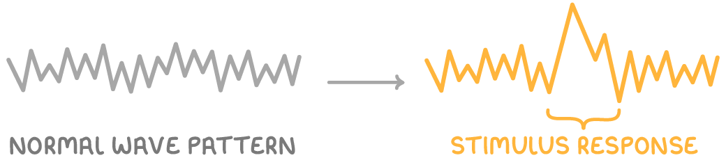
- Stimulus response - When a specific stimulus is presented, it causes a distinct alteration in the brain's electrical wave pattern, visible on an EEG.
- Whole brain activity - Like EEGs, ERPs reflect the activity of the entire brain rather than individual neurons.
Applications of ERPs
- Memory research - ERPs provide valuable data on how the brain processes and stores information.
- Psychiatric disorder studies - Research by Miltner et al. (2000) showed that individuals with phobias exhibit ERPs of higher amplitude when exposed to feared objects compared to non-phobic individuals.
Advantages of ERPs
- Non-invasive approach - ERPs can be studied without physical intrusion, similar to EEGs.
- Detailed stimulus insights - They offer precise information on brain reactions to specific triggers.
Limitations of ERPs
- Spatial resolution issues - Like EEGs, ERPs lack accuracy in locating the exact source of brain activity.
Post-mortem examinations for structural analysis
Post-mortem examinations involve the dissection of a deceased person's brain to study its internal structure. This method has historically been crucial for understanding brain anatomy and its relation to various conditions.
How post-mortem examinations are conducted
- Physical dissection - After a person has passed away, their brain is carefully examined and dissected to observe internal features and abnormalities.
Applications of post-mortem examinations
- Identifying structural abnormalities - These examinations can reveal physical changes linked to medical conditions. For instance, Brown et al. (1986) found enlarged ventricles in the brains of schizophrenia patients through post-mortem studies.
- Evidence for localisation of function - Historical research by Paul Broca used post-mortem analysis to identify Broca's area, linking damage in this region to speech production issues in patients.
Advantages of post-mortem examinations
- Detailed structural insights - They provide direct access to the brain's anatomy, offering information not always visible through scans.
Limitations of post-mortem examinations
- Post-death limitation - Examinations can only occur after death, meaning the individual cannot benefit from the findings.
- Causation challenges - While abnormalities can be observed, establishing cause and effect (e.g., whether enlarged ventricles cause schizophrenia) remains difficult with this method alone.