2.1 - Techniques for Studying the Brain
Functional magnetic resonance imaging (fMRI)
Functional magnetic resonance imaging (fMRI) is a technique that produces three-dimensional scans to reveal both the structure and activity of the brain. It detects real-time changes in brain function by tracking blood flow, offering insights into which areas become active during specific tasks.
How fMRI works
fMRI relies on a powerful magnetic field and radio waves to monitor blood oxygenation levels. Active brain regions require more oxygen and glucose, so oxygenated blood flows to these areas. The molecules in oxygenated blood react differently to the magnetic field compared to deoxygenated blood, allowing the scan to highlight active zones.
Applications and examples of fMRI
- Researching brain function and structure - Participants perform tasks inside the scanner, such as memory games, which activate relevant brain areas like those involved in recall.
- Diagnosing medical issues - fMRI identifies damaged or diseased brain tissue, aiding in the detection of conditions.
- Studying psychological disorders - Shergill et al (2001) used fMRI to examine brain activity during hallucinations in a patient with schizophrenia, revealing which regions were involved.
Strengths and limitations of fMRI
Strengths:
- Provides a non-invasive method to observe brain activity in real time.
Limitations:
- Equipment is costly to purchase and operate.
- Participants must remain completely still in a confined space, which can be difficult for those with claustrophobia.
- Offers poor temporal resolution, meaning it struggles to capture precise timing of brain changes.
Electroencephalogram (EEG)
An electroencephalogram (EEG) measures the brain's overall electrical activity by detecting signals from groups of neurons firing simultaneously. It uses electrodes placed on the scalp to record these patterns, which vary with states of arousal or consciousness.
How EEG works
Multiple electrodes on the scalp pick up electrical signals from synchronised neuron activity, producing wave patterns. These patterns differ across states, such as various sleep stages, where each stage shows distinct waveforms.
Applications and examples of EEG
- Sleep studies - EEG identifies wave patterns associated with different sleep phases.
- Investigating psychological conditions - It examines disorders like depression and schizophrenia.
- Diagnostic tool - EEG helps confirm conditions by revealing unusual electrical activity.
- Research evidence - A meta-analysis by Boutros et al (2008) found that people with schizophrenia exhibited unusual EEG wave patterns compared to healthy individuals.
Strengths and limitations of EEG
Strengths:
- Non-invasive and more affordable than fMRI.
- Provides good temporal resolution for tracking rapid changes in brain activity.
Limitations:
- Poor spatial resolution, making it challenging to pinpoint the exact brain regions generating the waves.
- Cannot detect activity from individual neurons, only from groups.
Event-related potentials (ERPs)
Event-related potentials (ERPs) are specific changes in EEG wave patterns triggered by particular stimuli. They reveal how the brain processes information in response to events, offering a refined view of cognitive reactions.
How ERPs work
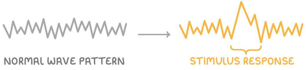
ERPs capture alterations in EEG waves when a stimulus is presented, with different stimuli producing unique waveform changes. This method isolates brain responses to specific inputs, enhancing the detail from standard EEG data.
Applications and examples of ERPs
- Memory and information processing research - ERPs are widely used to explore how the brain handles data, such as in recall tasks.
- Studying psychiatric disorders - Differences in ERPs appear in conditions like phobias.
- Research evidence - Miltner et al (2000) observed that individuals with phobias showed ERPs with higher amplitude when exposed to feared objects, compared to those without phobias.
Strengths and limitations of ERPs
- Strengths - Builds on EEG's temporal precision to link brain waves directly to stimuli.
- Limitations - Shares EEG's poor spatial resolution.
Post-mortem examinations
Post-mortem examinations involve analysing the brain after death to inspect its internal structure directly. This method allows detailed observation of physical features that scans might miss, contributing to understanding brain abnormalities.
How post-mortem examinations work
The brain is dissected post-death, enabling researchers to examine tissues and structures for irregularities that could explain behaviours or conditions observed during life.
Applications and examples of post-mortem examinations
- Identifying structural abnormalities - They reveal issues like enlarged brain ventricles linked to disorders.
- Evidence for localisation of function - Paul Broca's examinations of patients with speech difficulties identified damage in a consistent area, now known as Broca's area, responsible for speech production.
- Research evidence - Brown et al (1986) found enlarged ventricles in the brains of people with schizophrenia through post-mortem analysis.
Strengths and limitations of post-mortem examinations
Strengths:
- Provides direct access to brain tissue for precise structural insights.
Limitations:
- The individual cannot benefit from the findings, as the examination occurs after death.
- Cannot establish cause and effect; for example, abnormalities like enlarged ventricles in schizophrenia may not be caused by the disorder itself.
Overall evaluation of brain study methods
While these techniques offer valuable data on brain structure and function, they have shared challenges. Methods like fMRI, EEG, and ERPs provide non-invasive insights but vary in resolution, with fMRI excelling in spatial detail and EEG/ERPs in timing. Post-mortems give direct evidence but lack real-time applicability. Importantly, none definitively prove causation, as correlations (e.g., brain changes in disorders) do not confirm whether the changes cause the condition or result from it.