19.1 - Nuclear Magnetic Resonance Spectroscopy
- 1
What NMR spectroscopy is
- 2
Chemical environments and chemical shifts
- 3
The use of tetramethylsilane (TMS) as a reference standard
- 4
Interpreting 13C NMR spectra
NMR determines molecular structure
Nuclear magnetic resonance (NMR) spectroscopy is a powerful analytical technique that is used to understand the structure of molecules.
There are two main types:
- 13C NMR - This form provides information about the positions of carbon atoms within a molecule.
- 1H NMR - This form provides information about the positions of hydrogen atoms within a molecule.
The basis of NMR involves analysing the changes in magnetic properties of atomic nuclei brought about by their surrounding molecular environments.
Chemical environments influence magnetic properties
In NMR spectroscopy, when a molecule is placed in an external magnetic field, the nuclei within the molecule experience varying degrees of shielding from the external magnetic field. This shielding is due to their local "chemical environment" which encompasses all atoms, functional groups, and bonds directly connected to the nucleus.
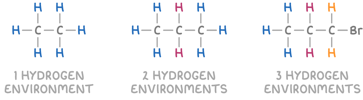
The electron density surrounding each nucleus acts as a magnetic shield, protecting it from the full strength of the external magnetic field. The degree of shielding varies depending on the specific chemical environment, with nuclei in different environments experiencing slightly different resonance frequencies when absorbing radio waves.
The more shielded a nucleus is, the lower its resonance frequency will be, as it requires less energy to flip its spin state in the presence of an external magnetic field.
By analysing these variations in resonance frequencies various nuclear environments, NMR spectroscopy provides valuable information about molecular structure.
Chemical shift
In NMR spectroscopy, chemical shift (δ) is a measure of the difference in resonant frequency between a given nucleus and a reference standard. It is expressed in parts per million (ppm) and it increases from right to left on the x-axis of an NMR spectrum.
Tetramethylsilane (Si(CH3)4), or TMS, is used as the standard in NMR spectroscopy because:
- TMS produces a single absorption peak due to all its carbon and hydrogen atoms being in identical chemical environments.
- The TMS peak appears at a lower frequency, located to the right of most analytes on the NMR spectrum, making it an ideal reference point.
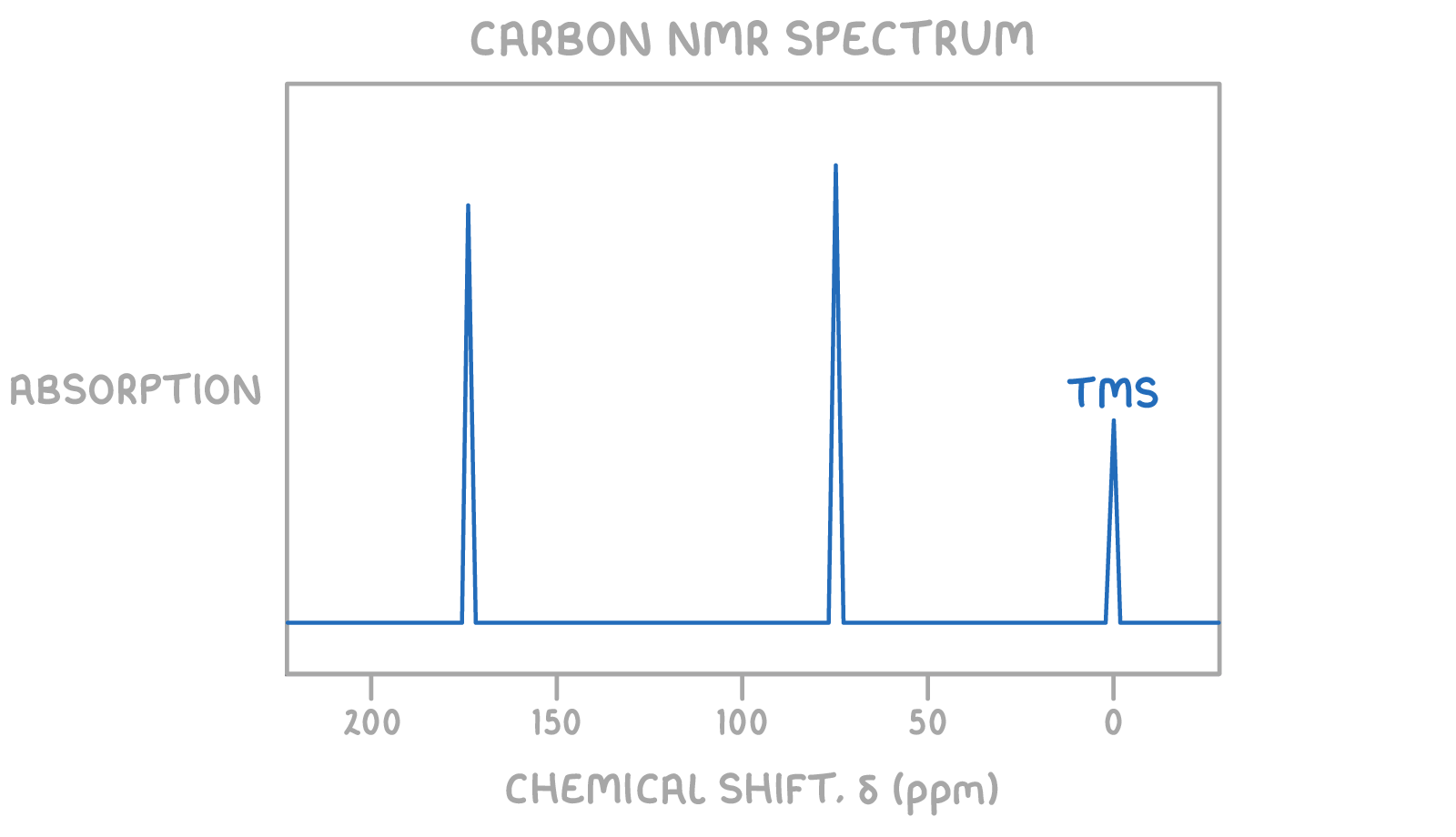
The TMS peak is assigned a chemical shift value of 0 ppm, serving as the reference point for measuring chemical shifts of other peaks in the spectrum.
Peaks from the sample that absorb at frequencies higher than that of TMS appear at higher chemical shift values measured relative to the TMS peak at 0 ppm. These peaks are located to the left of the TMS peak, indicating that the corresponding nuclei are less shielded than TMS. Consequently, these nuclei experience a stronger effective magnetic field.
Number of peaks shows distinct carbon environments
The number of peaks present on a 13C NMR spectrum indicates the number of magnetically distinct carbon environments in the molecule.
Each unique carbon environment produces its own peak, meaning that each peak represents a set of equivalent carbon atoms.
For example, chloroethane (CH3CH2Cl) has two magnetically different carbon environments and thus two peaks in its 13C NMR spectrum:
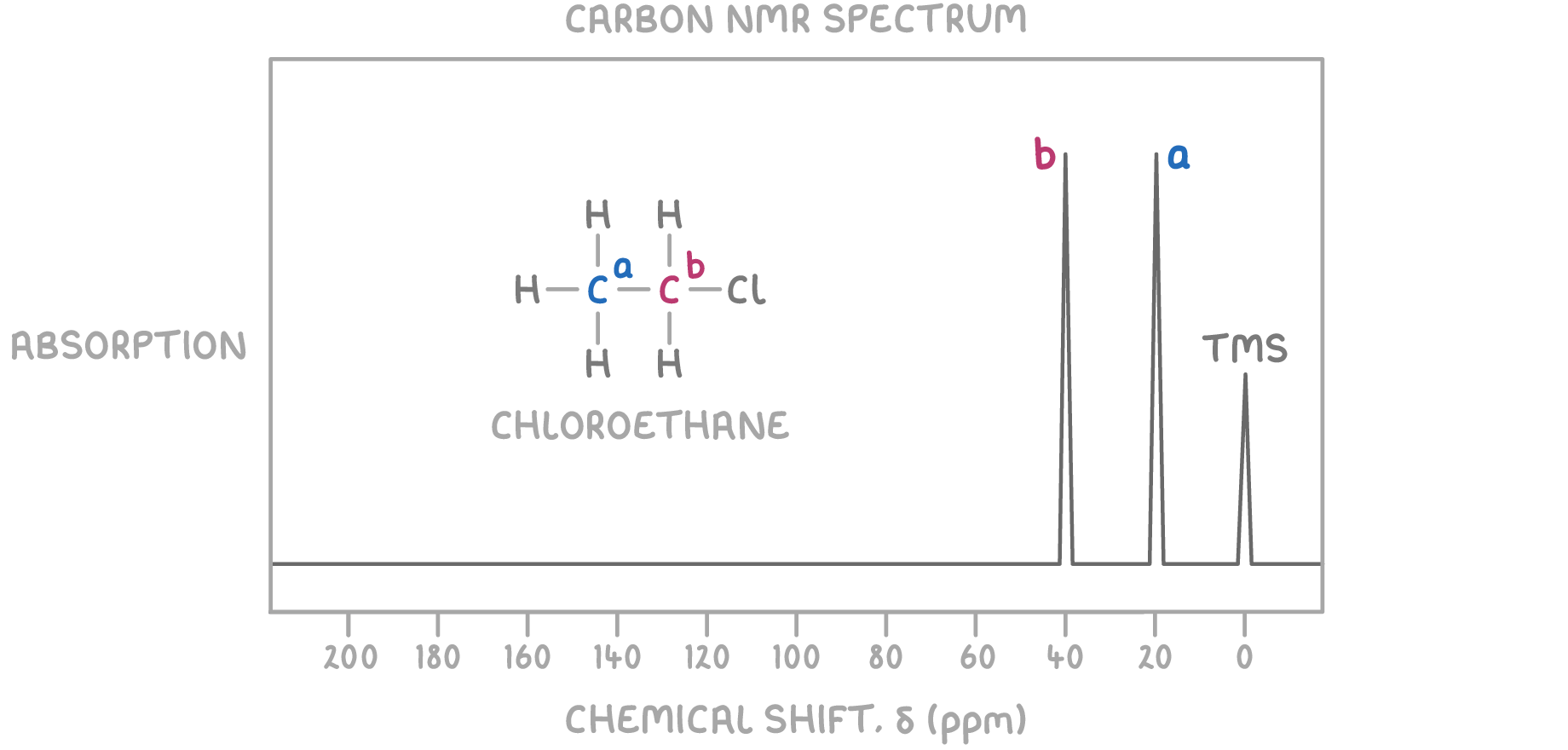
The differing bonding of chlorine and hydrogen atoms to each carbon alters the electron density around each carbon, making their environments distinguishable.
Even molecules with multiple carbons, like 1,3-dibromocyclohexane, have a predictable number of peaks based on symmetry:
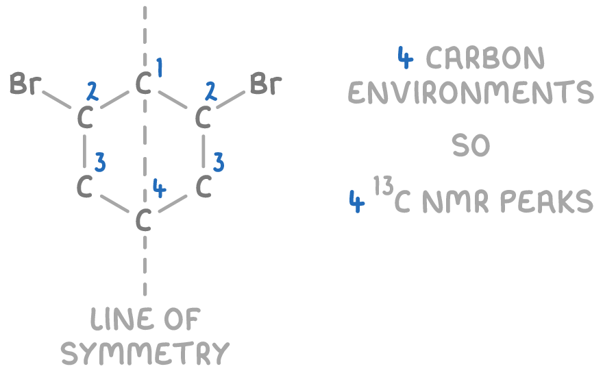
Thus, by carefully examining a molecule’s structure, the number of expected 13C NMR peaks can be determined.
Using chemical shift data tables
Chemical shift values offer insights into the functional groups attached to carbon nuclei.
Data tables compile known chemical shift ranges for different carbon environments relative to TMS:
| Chemical shift, δ (ppm) | Type of carbon |
|---|---|
| 5 – 40 | C–C |
| 10 – 70 | R–C–Cl or R–C–Br |
| 20 – 50 | R–C(=O)–C |
| 25 – 60 | R–C–N (amines) |
| 50 – 90 | C–O (alcohols, ethers or esters) |
| 90 – 150 | C=C (alkenes) |
| 110 – 125 | R–C≡N (nitrile) |
| 110 – 160 | Aromatic |
| 160 – 185 | R–C=O (ester or carboxylic acid) |
| 190 – 220 | R–C=O (aldehyde or ketone) |
Matching peaks from a spectrum to table entries allows carbon environments to be identified.
However:
- Overlapping shift ranges mean that assignments may not be definitive.
- Additional evidence is needed to confirm environment identities.
For example, a peak at ~30 ppm could represent C-C, C-Cl, or C-Br environments based on the table.
Determining molecular structures
Combining evidence from the number of peaks, chemical shift values, and molecular formula allows unknown structures to be deduced.
This methodical approach involves the following steps:
- Count the number of distinct peaks to understand the variety of carbon environments.
- Use the chemical shift data to hypothesise about the types of carbon present.
- Combine all available evidence to propose a molecular structure that fits the given data and the molecular formula.
Worked example 1 - Identifying the structure of an unknown molecule
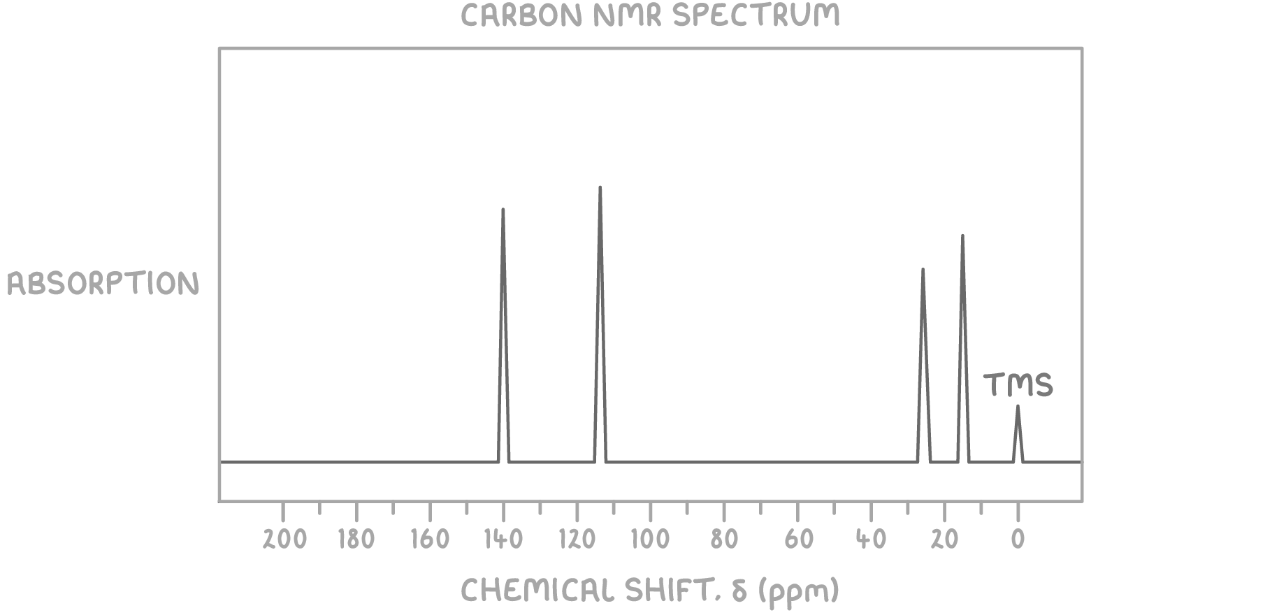
Using the molecular formula C4H8 and the 13C NMR spectrum provided below, determine the structure of the unknown molecule.
Step 1: Count peaks
The presence of 4 peaks indicates there are 4 magnetically distinct carbon environments in the molecule.
Step 2: Deduce environments from chemical shifts
The peaks at δ 113 ppm and δ 140 ppm suggest carbon atoms involved in a C=C bond, typical for alkenes.
The peaks at δ 13 ppm and δ 27 ppm suggest various types of saturated C-C bond environments, likely representing methyl groups.
Step 3: Consider the molecular formula
Given the molecular formula C4H8, the compound is unsaturated, indicating the presence of a double bond due to the hydrogen count being less than the maximum for a four-carbon alkane (C4H10). This confirms the alkene nature of the compound.
Step 4: Match environments to potential structures
Considering the presence of a double bond indicated by the peaks at δ 113 ppm and δ 140 ppm:
- A straight-chain alkene is required to match the number of carbon peaks observed.
- But-1-ene fits all criteria: it has a double bond creating distinct environments for the carbon atoms (at δ 113 ppm and δ 140 ppm), and two distinct types of C-C environments corresponding to the peaks at δ 13 ppm and δ 27 ppm.
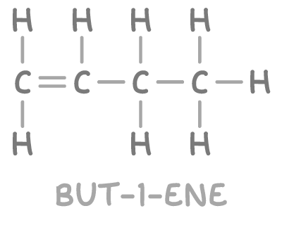
Therefore, the unknown molecule is identified as but-1-ene.
Worked example 2 - Identifying the structure of an unknown molecule
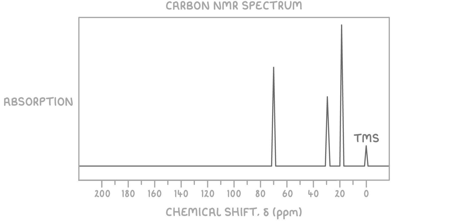
Using the molecular formula C4H10O and the 13C NMR spectrum provided below, determine the structure of the unknown molecule.
Step 1: Count peaks
The presence of 3 peaks indicates there are 3 magnetically distinct carbon environments in the molecule.
Step 2: Deduce environments from chemical shifts
The peak at δ 70 ppm suggests a C-O bond, typical for alcohols.
The peaks at δ 19 ppm, and δ 31 ppm suggest various types of C-C bond environments, indicating a chain with branching or differing degrees of saturation.
Step 3: Consider the molecular formula
Given the molecular formula C4H10O, the compound is saturated, indicating the absence of double or triple bonds. The presence of oxygen and the saturation level suggest an alcohol.
Step 4: Match environments to potential structures
Considering the alcohol functional group suggested by the δ 70 ppm peak:
- A straight-chain alcohol would not adequately account for the diversity of the chemical shift values observed.
- A molecule like 2-methylpropan-1-ol fits all criteria: it has an C-OH alcohol group at δ 70 ppm (carbon c) and two distinct types of C-C environments corresponding to the peaks at δ 19 ppm and δ 31 ppm.
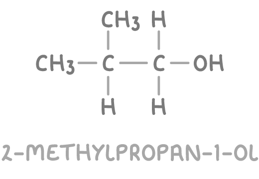
Therefore, the unknown molecule is identified as 2-methylpropan-1-ol.