8.8 - How the Heartbeat is Controlled
- 1
How the heart beat is controlled
- 2
How electrocardiograms monitor heart function
How the heart beat is controlled
Cardiac muscle is myogenic, which means that the contraction of cardiac muscle initiates within the heart itself. The basic rhythm of the heart is maintained by a wave of electrical excitation.
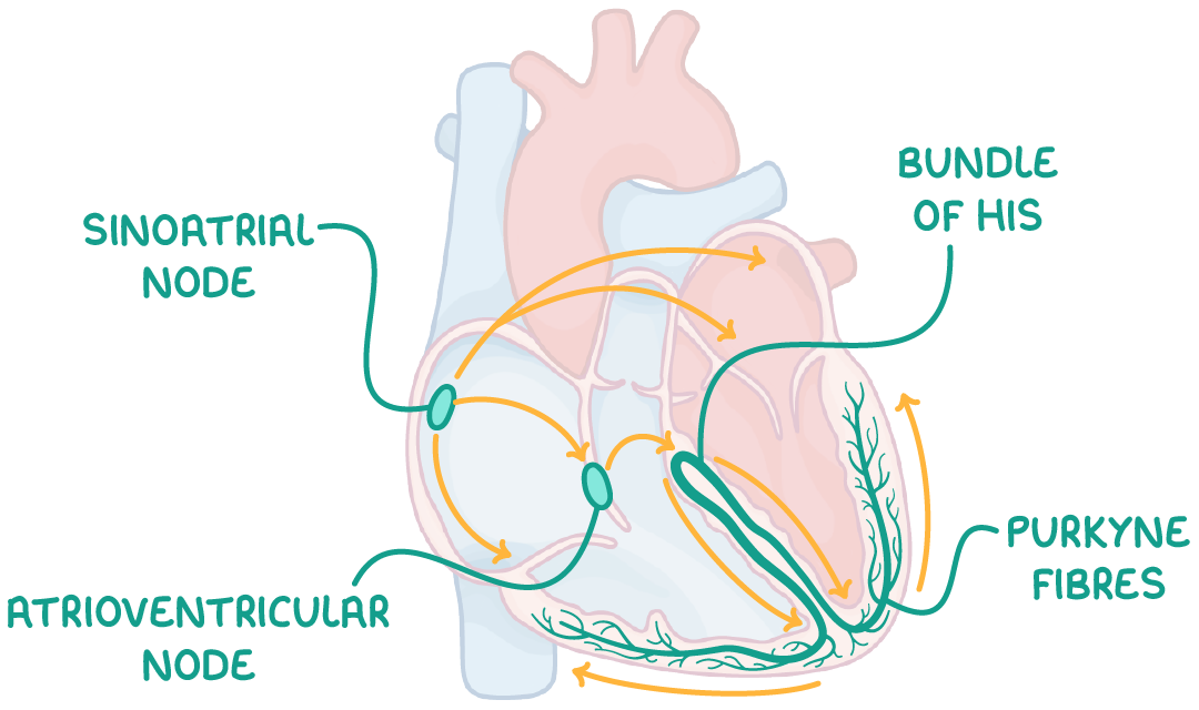
This wave travels through the following structures in this order:
- Sinoatrial node (SAN) - Initiates the heart beat by stimulating the atria to contract.
- A layer of collagen fibres - Prevents direct electrical flow from atria to ventricles.
- Atrioventricular node (AVN) - Picks up the electrical activity from the SAN and imposes a slight delay.
- Bundle of His - Receives electrical activity from the AVN and conducts the wave of excitation to the apex (base) of the heart.
- Purkyne fibres - These branch off the bundle of His, causing the right and left ventricles to contract from the bottom upwards.
How electrocardiograms monitor heart function
Electrocardiograms (ECGs) record the heart's electrical activity using electrodes.
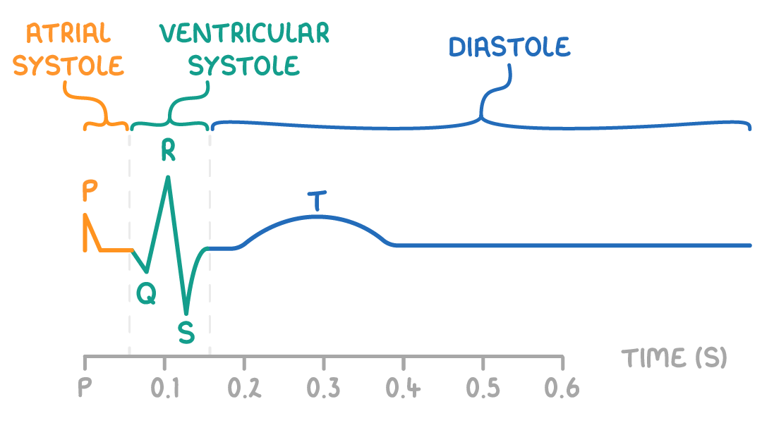
An ECG shows the various stages of the cardiac cycle:
- P wave - Atrial systole
- QRS - Ventricular systole
- T wave - Diastole
ECGs help diagnose abnormalities like:
- Tachycardia - This is an abnormally rapid heart rate.
- Bradycardia - This is an abnormally slow heart rate.
- Ectopic heartbeats - This is extra heartbeats out of the normal rhythm.
- Atrial fibrillation - This is the abnormally rapid and ineffective contraction of the atria.
9.1 - Xylem & PhloemNext up
How were these notes?