19.6 - Microarrays
- 1What microarrays are and how they work
- 2Using microarrays to compare genes in different species
- 3Using microarrays to identify gene expression
What is a microarray?
A microarray, also known as a gene chip or DNA chip, is a small glass or plastic slide printed with thousands of DNA sequences at specific locations.
These sequences act as probes to detect complementary DNA or RNA strands, allowing the rapid analysis of many genes simultaneously.
How microarrays work
Microarrays involve hybridisation, which occurs when two single-stranded DNA molecules with complementary base pairs join to form double-stranded DNA.
How microarrays work:
- Multiple copies of short, single-stranded DNA probes are fixed to specific spots on the chip, with each spot containing multiple copies of a single probe.
- DNA or RNA samples, tagged with fluorescent markers, are washed over the chip.
- Complementary base pairing allows the samples to bind (hybridise) with matching probes.
- Any strands that do not bind are washed away.
- Scanners equipped with ultraviolet light detect and visualise hybridised probes by their fluorescent tags.
- The data is then analysed by computers to identify the presence of genes and their expression levels.
A single microarray can have probes for an entire organism's genome, offering a comprehensive genetic analysis tool.
Using microarrays to compare genes in different species
Microarrays allow the comparison of genetic material between the genomes of different species.
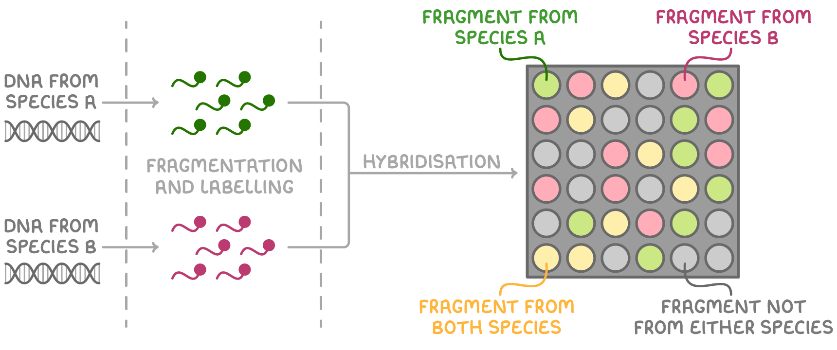
The process involves:
- Extracting DNA from the species of interest.
- Fragmenting this DNA and labelling each species' DNA with distinct fluorescent tags (for example, green for species A and red for species B).
- Mixing and hybridising the labelled DNA to the microarray probes.
- Scanning the microarray to detect hybridised DNA.
- The appearance of coloured spots shows that a DNA fragment has successfully hybridised with complementary DNA probes.
The colour of the microarray spots reveals the origin of the DNA fragments, with specific colours indicating specific species.
| Colour of microarray spot | Which species the DNA fragment is from | What this indicates |
|---|---|---|
| The colour of the fluorescent label for a particular species | The species indicated by the colour of the spot | The DNA fragment has hybridised with the probes, indicating the presence of that species' DNA |
| A colour that was not used for labelling | Both species | Both species have the same DNA sequence for that fragment, implying a common gene between the two species |
| No colour | Neither species | No DNA hybridised with the probes, indicating that a particular gene is not present in either species |
Using microarrays to identify gene expression
Microarrays are also pivotal in determining which genes are active within cells. This process is particularly useful for contrasting gene expression between cancerous and non-cancerous cells to pinpoint genes that are active in cancer.
The process involves:
- Isolating mRNA from cell samples.
- Using reverse transcriptase to convert mRNA into complementary DNA (cDNA), which is then fluorescently labelled.
- Using PCR to increase the amount of cDNA if the initial quantity of mRNA is low.
- Hybridising the labelled cDNA to the microarray probes.
- Scanning the microarray to detect the cDNA that has bound.
- The intensity of the spots on the microarray correlates with gene expression levels; brighter spots indicate higher gene activity due to the greater presence of mRNA.