Microscopy - exam practice
Microscopy
Question 1 of 9
The image below shows part of a cell observed using a microscope.
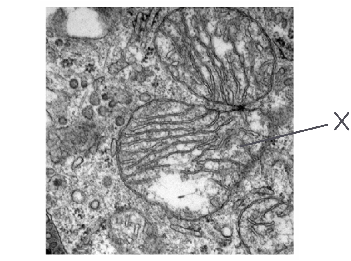
Name the organelle labelled X in the diagram above.
Which type of microscope was used to obtain this image? Explain your answer.
Describe what is meant by the term resolution.
Compare the resolutions obtained by a scanning electron microscope and a transmission electron microscope.
Include the differences between the images produced by each microscope.
A student wanted to investigate the chloroplasts inside a plant cell.
The image below shows part of the plant Calypogeia neesiana as seen using an optical microscope.
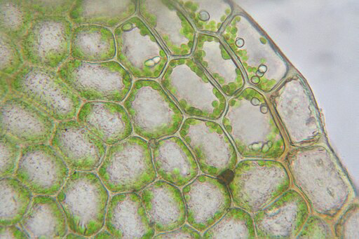
Describe how the student would have used the optical microscope to observe these cells using a high power lens.
Explain why the structures inside the chloroplasts cannot be seen in the image above.
A transmission electron microscope would enable the student to the chloroplasts in more detail.
Describe how a transmission electron microscope works to give a higher image resolution.
Describe the limitations of using a transmission electron microscope.
A student wanted to investigate the process of cell division.
He wasn't sure whether it was best to use a light microscope or an electron microscope.
Suggest the advantages of using a light microscope rather than an electron microscope to study cell division.
The student suggested that he could use a light microscope to investigate cell division while also observing the structure of mitochondria.
Explain why the student is not correct.
An electron microscope would give a higher resolution.
Explain the difference between magnification and resolution.
The student decided to investigate cell division using a light microscope. He used a stain called toludine blue when preparing his slides.
Describe the advantages of staining specimens before viewing them under a microscope.
A student wanted to investigate the number of chloroplasts in some plant cells.
Describe how he could make a temporary mount of a piece of plant tissue to observe the number of chloroplasts in the cells when using an optical microscope.
Describe how the student could use the temporary mounts to determine the mean number of chloroplasts in the leaf cells.
State two features of a plant cell that would not be visible using medium power of an optical microscope.
Compare the advantages and disadvantages of optical microscopes and transmission electron microscopes.
The diagram below shows an image of part of a eukaryotic cell as viewed under a microscope.
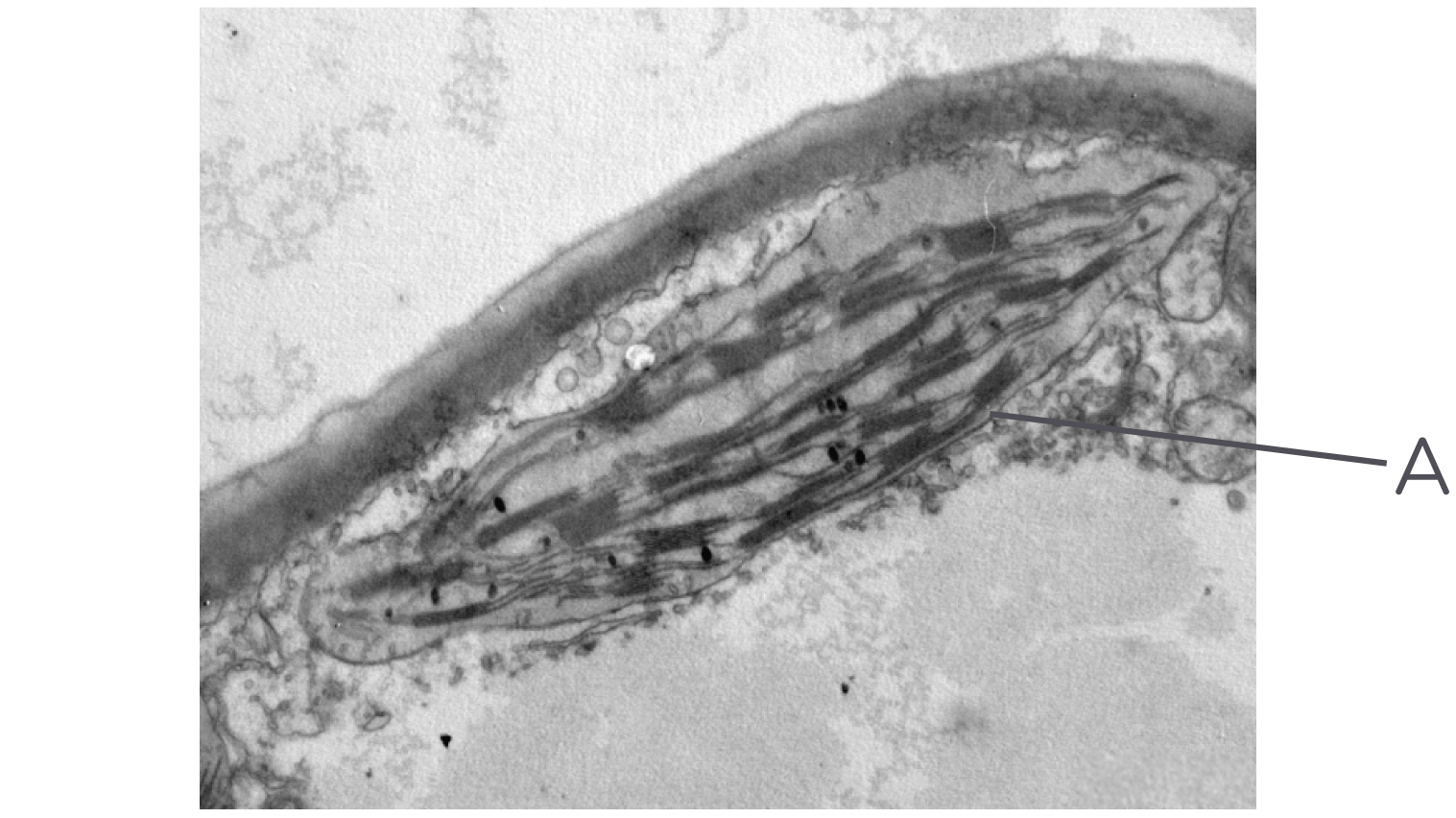
Name organelle A shown in the diagram above.
Describe the function of organelle A.
The actual size of this organelle is 3.5 µm, whereas its size on the image is 4 mm.
Calculate the magnification of this image. Give your answer to the nearest whole number.
Give one piece of evidence to support that the cell shown above was observed using an electron microscope.
The diagram below shows a breast cancer cell as seen through an electron microscope.
Included in the diagram is a graticule in which the markings are 0.1 mm apart.
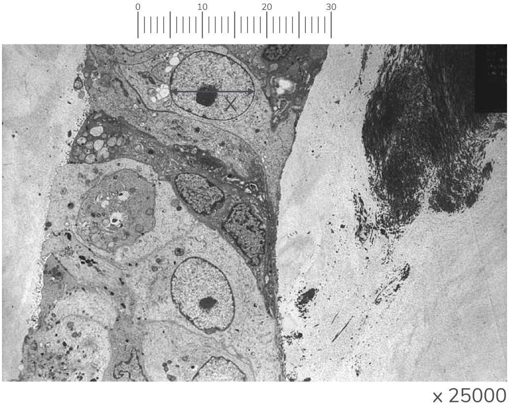
Use the graticule to calculate the actual width of the cell labelled X in the diagram above. Give your answer in µm.
Explain why a transmission electron microscope rather than an optical microscope was used to study the cancer cell.
Describe one advantage of using a transmission electron microscope rather than a scanning electron microscope.
Describe one advantage of using a scanning electron microscope rather than a transmission electron microscope.
The diagram below shows some cancer cells when viewed under a microscope.
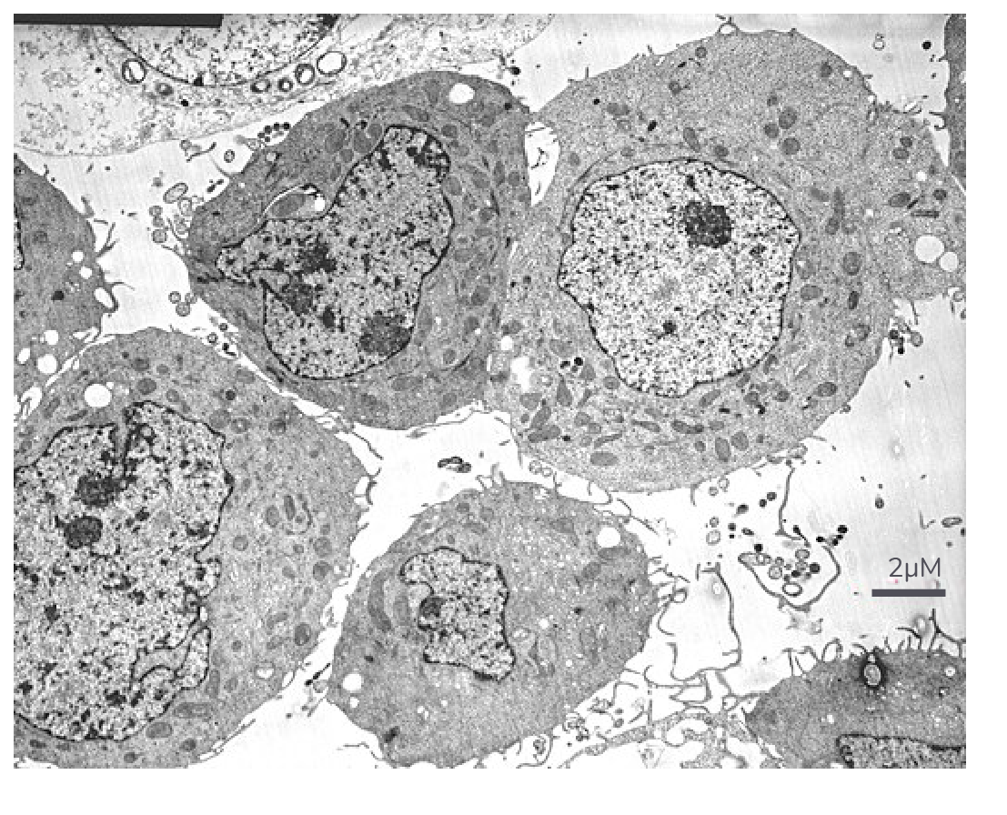
Which kind of microscope was used to obtain this image? Explain your answer.
The image size of the scale bar is 4 mm. Calculate the magnification of the image.
State the resolution that can be achieved using an optical microscope and a transmission electron microscope.
Another type of electron microscope is the scanning electron microscope.
Compare the benefits and limitations of transmission electron microscopes and scanning electron microscopes.
The diagram below shows an electron micrograph of the bacterium Gemmata obscuriglobus.
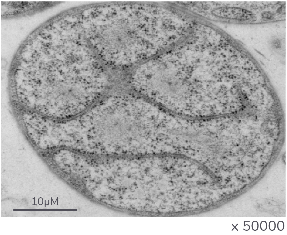
The scale bar shown on the diagram represents a measurement of 10 µm.
Calculate the image size of this scale bar.
Explain why it is possible to see the structure of a prokaryotic cell with an electron microscope but not with an optical microscope.
Describe and explain how cell fractionation and ultracentrifugation can be used to isolate ribosomes from a suspension of bacterial cells.
A student prepared a sample of mitochondria from a sample of muscle tissue. She used the following method:
- Add the muscle tissue to an ice-cold, buffered and isotonic solution.
- Mix the muscle and solution in a blender.
- Filter the mixture.
- Spin the filtered liquid at low speed in a centrifuge. A pellet will appear at the bottom of the tube.
- Pour off the liquid above the pellet into a second tube and spin at a higher speed.
Explain why the solution has to be ice-cold, buffered, and isotonic.
Explain why the solution was blended.
Explain why the solution was filtered after blending.
Name the organelle you would expect to find to make up most of the first pellet.
Explain why the second centrifuge tube was spun at a higher speed.